. 2017 Nov 24;292(47):19441-19457.
doi: 10.1074/jbc.M117.815910. Epub 2017 Oct 11.

The molecular mechanism of N-acetylglucosamine side-chain attachment to the Lancefield group A carbohydrate in Streptococcus pyogenes

Affiliations

The molecular mechanism of N-acetylglucosamine side-chain attachment to the Lancefield group A carbohydrate in Streptococcus pyogenes

Jeffrey S Rush et al. J Biol Chem. .

Abstract

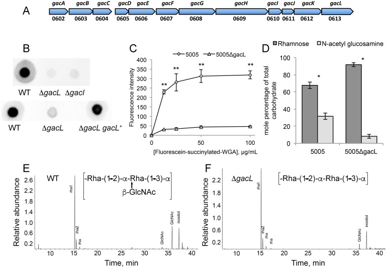
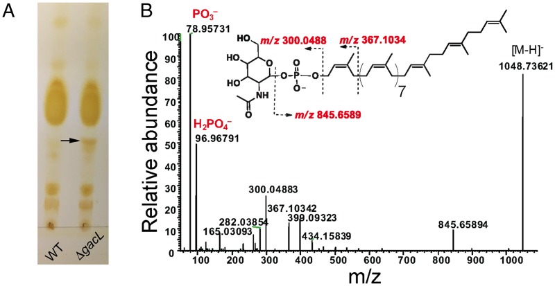
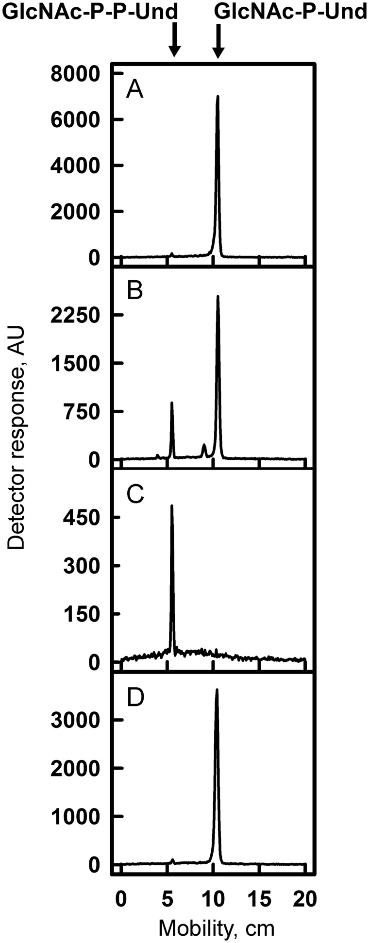
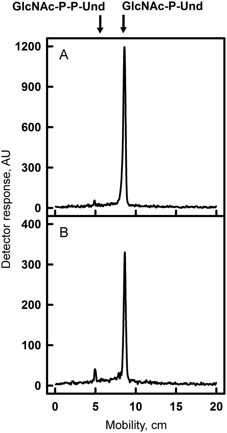
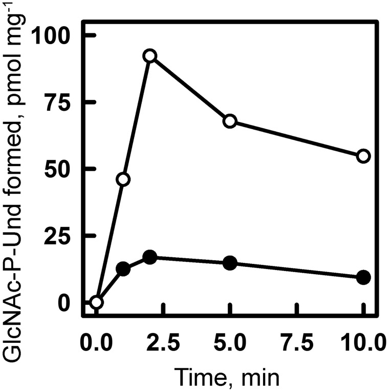
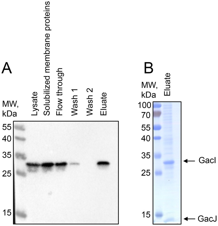
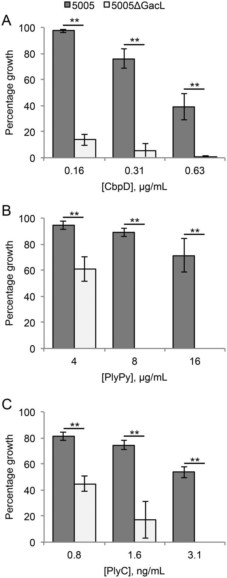
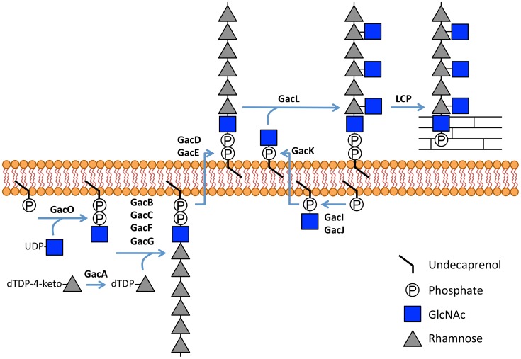
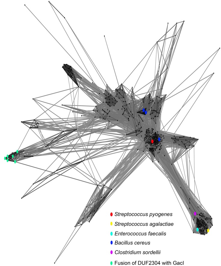
In many Lactobacillales species (i.e. lactic acid bacteria), peptidoglycan is decorated by polyrhamnose polysaccharides that are critical for cell envelope integrity and cell shape and also represent key antigenic determinants. Despite the biological importance of these polysaccharides, their biosynthetic pathways have received limited attention. The important human pathogen, Streptococcus pyogenes, synthesizes a key antigenic surface polymer, the Lancefield group A carbohydrate (GAC). GAC is covalently attached to peptidoglycan and consists of a polyrhamnose polymer, with N-acetylglucosamine (GlcNAc) side chains, which is an essential virulence determinant. The molecular details of the mechanism of polyrhamnose modification with GlcNAc are currently unknown. In this report, using molecular genetics, analytical chemistry, and mass spectrometry analysis, we demonstrated that GAC biosynthesis requires two distinct undecaprenol-linked GlcNAc-lipid intermediates: GlcNAc-pyrophosphoryl-undecaprenol (GlcNAc-P-P-Und) produced by the GlcNAc-phosphate transferase GacO and GlcNAc-phosphate-undecaprenol (GlcNAc-P-Und) produced by the glycosyltransferase GacI. Further investigations revealed that the GAC polyrhamnose backbone is assembled on GlcNAc-P-P-Und. Our results also suggested that a GT-C glycosyltransferase, GacL, transfers GlcNAc from GlcNAc-P-Und to polyrhamnose. Moreover, GacJ, a small membrane-associated protein, formed a complex with GacI and significantly stimulated its catalytic activity. Of note, we observed that GacI homologs perform a similar function in Streptococcus agalactiae and Enterococcus faecalis In conclusion, the elucidation of GAC biosynthesis in S. pyogenes reported here enhances our understanding of how other Gram-positive bacteria produce essential components of their cell wall.

Keywords: Streptococcus pyogenes (S. pyogenes); carbohydrate biosynthesis; cell wall; glycosyltransferase; lipid intermediate; polysaccharide.


Conflict of interest statement

The authors declare that they have no conflicts of interest with the contents of this article

Figures

Figure 1.
Figure 1.
Map of S. pyogenes genes involved in GAC biosynthesis and analysis of 5005ΔgacI and 5005ΔgacL deletion mutants. A, GAC biosynthesis gene cluster. Numbers below represent MGAS5005 gene designations. B, representative immunoblot analysis of cell-wall fractions isolated from MGAS5005, 5005ΔgacI, 5005ΔgacL, and 5005ΔgacL gacL+. Data are representative of biological triplicates. C, binding of N-acetylglucosamine-specific fluorescein-succinylated WGA to whole MGAS5005 and 5005ΔgacL was measured. Data are the average of three replicates ± S.D. D, rhamnose and GlcNAc mole percentage of total carbohydrate was determined by GC-MS for cell wall material isolated from MGAS5005 and 5005ΔgacL following methanolysis as described under “Experimental procedures.” Data are the average of four replicates ± S.D. The asterisks indicate statistically different values (*, p < 0.05; **, p < 0.01) as determined by the Student's t test. E and F, GC-MS chromatograms for glycosyl composition analysis of cell wall isolated MGAS5005 and 5005ΔgacL. The deduced schematic structure of the repeating unit of GAC is shown for each strain. The chromatograms are representative of four separate analyses performed on two different cell wall preparations.
Figure 2.
Figure 2.
Purification and identification of GlcNAc-phosphate-undecaprenol in 5005ΔgacL. A, TLC analysis of phospholipids isolated from MGAS5005 and 5005ΔgacL. Phospholipids extracted from bacterial strains were separated by TLC on Silica Gel G in CHCl3/CH3OH/H2O/NH4OH (65:25:4:1). Position of the novel, alkaline-resistant, and acid-labile phospholipid accumulating in 5005ΔgacL is indicated by the arrow. The results are representative of three separate experiments. B, ESI-MS/MS analysis of the novel phospholipid isolated from 5005ΔgacL. The spectrum is assigned to GlcNAc-phosphate-undecaprenol.
Figure 3.
Figure 3.
TLC of [3H]GlcNAc-lipids from in vitro incubations of GAS mutants and B. cereus membranes with UDP-[3H]GlcNAc. Membrane fractions from MGAS5005 (A), B. cereus (B), 5005ΔgacI (C), or 5005ΔgacL (D) were incubated with UDP-[3H]GlcNAc and analyzed for [3H]GlcNAc-lipid synthesis by TLC. Reaction mixtures contained 50 mm Tris-Cl, pH 7.4, 5 mm 2-mercaptoethanol, 20 mm MgCl2, 1 mm ATP, 5 μm UDP-[3H]GlcNAc (486 cpm/pmol), and bacterial membrane suspension (100–200 μg of membrane protein) in a total volume of 0.02 ml. Following a 10-min pre-incubation at 30 °C, GlcNAc-lipid synthesis was initiated by the addition of UDP-[3H]GlcNAc. After 10 min, reactions were processed for GlcNAc-lipid synthesis as described under “Experimental procedures.” The organic layers were dried and dissolved in a small volume of CHCl3/CH3OH (2:1), and a portion was removed and assayed for radioactivity by liquid scintillation spectrometry. The remainder was spotted on 10 × 20-cm plate of Silica Gel G and developed in CHCl3/CH3OH/NH4OH/H2O (65:25:1:4). [3H]GlcNAc-lipids were detected by scanning with an AR2000 Bioscan Radiochromatoscanner. AU, arbitrary units. The results are representative of three separate experiments.
Figure 4.
Figure 4.
TLC of [3H]GlcNAc-lipids from in vitro incubations of GAS and GBS membranes with UDP-[3H]GlcNAc. Membrane fractions from WT MGAS5005 (A) or GBS COH1 (B) were incubated with UDP-[3H]GlcNAc and analyzed for [3H]GlcNAc-lipid synthesis by TLC. Reaction mixtures were exactly as described in the legend to Fig. 3. After 5 min of incubation with UDP-[3H]GlcNAc, reactions were processed for GlcNAc-lipid synthesis as described under “Experimental procedures.” The organic layers were dried and dissolved in a small volume of CHCl3/CH3OH (2:1), and a portion was removed and assayed for radioactivity by liquid scintillation spectrometry. The remainder was spotted on 10 × 20-cm plate of Silica Gel G and developed in CHCl3/CH3OH/NH4OH/H2O (65:25:1:4). [3H]GlcNAc-lipids were detected by scanning with an AR2000 Bioscan Radiochromatoscanner. AU, arbitrary units. The results are representative of three separate experiments.
Figure 5.
Figure 5.
Effect of ATP on GlcNAc-P-Und synthesis in vitro in MGAS5005 membrane fractions. GlcNAc-P-Und synthesis in MGAS5005 membranes was assayed after the indicated time at 30 °C in the presence (○) or absence (●) of 1 mm ATP. Reaction mixtures were identical to those described in Fig. 3 except for the presence of ATP. Following incubation, incorporation into [3H]GlcNAc-P-Und was determined as described under “Experimental procedures.” The results are representative of three separate experiments.
Figure 6.
Figure 6.
GacI and GacJ exist as a detergent-stable complex in the membrane. GacJ and His-tagged GacI were co-expressed in E. coli Rosetta DE3 cells and extracted from the membrane fraction in 2.5% CHAPS. The proteins were purified using Ni-NTA-agarose in the presence of 2.5% CHAPS. A, fractions collected during Ni-NTA purification were analyzed by immunoblot using anti-His antibodies. B, eluted proteins were analyzed by SDS-PAGE. The results are representative of three separate experiments.
Figure 7.
Figure 7.
Analysis of GacO function in MGAS5005. A, GacO catalyzes the synthesis of GlcNAc-P-P-Und in E. coli membranes. Reaction mixtures contained 50 mm Tris-HCl, pH 7.4, 5 mm 2-mercaptoethanol, 20 mm MgCl2, 0.5% CHAPS, 20 μm Und-P (dispersed by ultrasonication in 1% CHAPS), 5 μm UDP-GlcNAc (452 cpm/pmol), and E. coli membrane fraction from either PR4019, CLM37, or CLM37:GacO strains. Following incubation for 10 min at 30 °C, incorporation of [3H]GlcNAc into [3H]GlcNAc-P-P-Und was determined as described under “Experimental procedures.” Data are the average of three replicates ± S.D. B and C, GlcNAc-P-P-Und can function as an acceptor substrate for rhamnosylation to form rhamnosyl-GlcNAc-P-P-Und in 5005ΔgacI membranes. Incubation conditions were as described in the legend to Fig. 3. B, after 35 min of incubation with UDP-[3H]GlcNAc, the reactions were analyzed for formation of [3H]GlcNAc lipids as described in Fig. 3. C, after 5 min of incubation with UDP-[3H]GlcNAc, the reactions were incubated with 20 μm TDP-rhamnose for an additional 30 min and analyzed for formation of [3H]GlcNAc lipids. The results are representative of three separate experiments. D, ESI-MS/MS spectrum of rhamnosyl-GlcNAc-P-P-Und formed during chase with TDP-rhamnose from GlcNAc-P-P-Und synthesized in situ in membranes from S. pyogenes 5005ΔgacI.
Figure 8.
Figure 8.
Absence of GacL increases sensitivity to cell wall amidases. Mid-exponential phase MGAS5005 and 5005ΔgacL were grown in the indicated concentrations of CbpD (A), PlyPy (B), and PlyC (C). The change in growth is represented as a percentage of growth where no amidase was present. Data are the average of three replicates ± S.D. The asterisk indicate statistically different values (**, p < 0.01) as determined by the Student's t test.
Figure 9.
Figure 9.
Schematic diagram of GAC biosynthesis. GAC is anchored to peptidoglycan presumably via phosphodiester bond. GAC biosynthesis is initiated on the inner leaflet of the plasma membrane where GacO produces GlcNAc-P-P-Und, which serves as a membrane-anchored acceptor for polyrhamnose synthesis catalyzed by the GacB, GacC, GacF, and GacG rhamnosyltransferases. Following polymerization, polyrhamnose is transferred to the outer leaflet of the membrane presumably by the GacD/GacE ABC transporter. Also in the inner leaflet of the membrane, GacI aided by GacJ produces GlcNAc-P-Und, which then diffuses across the plasma membrane to the outer leaflet aided by GacK. Subsequently, GacL transfers GlcNAc to polyrhamnose using GlcNAc-P-Und as glycosyl donor. Finally, protein members of LytR-CpsA-Psr phosphotransferase family presumably attach GAC to peptidoglycan. Several details of this biosynthetic scheme are still speculative, and further research will be required to definitively confirm this hypothetical pathway, but the overall organization is consistent with other isoprenol-mediated capsular polysaccharide pathways.
Figure 10.
Figure 10.
Sequence relationship of GacI family of proteins. Homology between GacI homologs is graphically displayed using CLANS analysis (37). Dots correspond to individual protein sequences selected as described under “Experimental procedures” and provided as supplemental file S1. Selected homologs of S. pyogenes GacI are highlighted by colored dots.

References

    1. Mistou M. Y., Sutcliffe I. C., and van Sorge N. M. (2016) Bacterial glycobiology: rhamnose-containing cell wall polysaccharides in Gram-positive bacteria. FEMS Microbiol. Rev. 40, 464–479 - PMC - PubMed
    1. Carapetis J. R., Steer A. C., Mulholland E. K., and Weber M. (2005) The global burden of group A streptococcal diseases. Lancet Infect. Dis. 5, 685–694 - PubMed
    1. McCarty M. (1952) The lysis of group A hemolytic streptococci by extracellular enzymes of Streptomyces albus. II. Nature of the cellular substrate attacked by the lytic enzymes. J. Exp. Med. 96, 569–580 - PMC - PubMed
    1. Lancefield R. C. (1933) A serological differentiation of human and other groups of hemolytic streptococci. J. Exp. Med. 57, 571–595 - PMC - PubMed
    1. van Sorge N. M., Cole J. N., Kuipers K., Henningham A., Aziz R. K., Kasirer-Friede A., Lin L., Berends E. T. M., Davies M. R., Dougan G., Zhang F., Dahesh S., Shaw L., Gin J., Cunningham M., et al. (2014) The classical lancefield antigen of group a Streptococcus is a virulence determinant with implications for vaccine design. Cell Host Microbe 15, 729–740 - PMC - PubMed

Publication types

MeSH terms

LinkOut - more resources