. 2017 Nov 10;121(11):1251-1262.
doi: 10.1161/CIRCRESAHA.117.311819. Epub 2017 Oct 11.

Lactate Promotes Synthetic Phenotype in Vascular Smooth Muscle Cells

Affiliations

Lactate Promotes Synthetic Phenotype in Vascular Smooth Muscle Cells

Libang Yang et al. Circ Res. .

Abstract

Rationale: The phenotypes of vascular smooth muscle cells (vSMCs) comprise a continuum bounded by predominantly contractile and synthetic cells. Some evidence suggests that contractile vSMCs can assume a more synthetic phenotype in response to ischemic injury, but the mechanisms that activate this phenotypic switch are poorly understood.

Objective: To determine whether lactate, which increases in response to regional ischemia, may promote the synthetic phenotype in vSMCs.

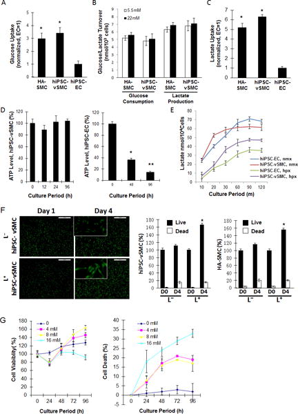
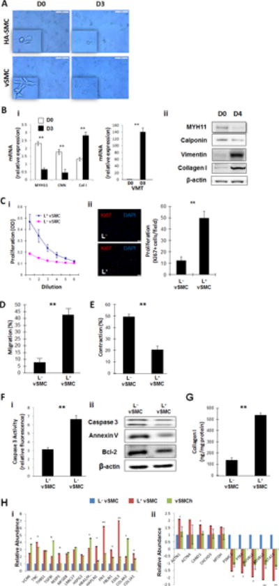
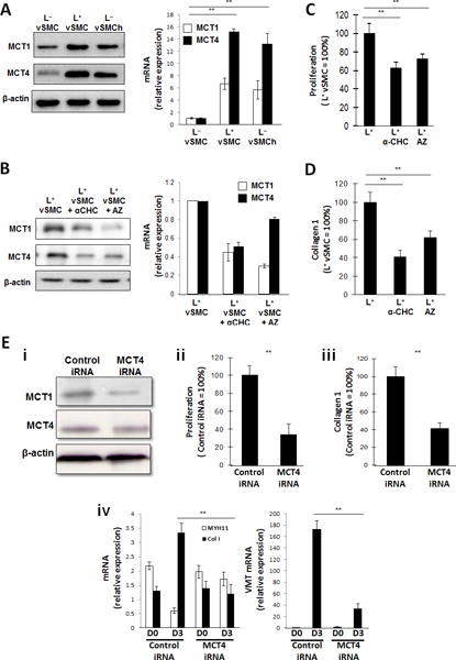
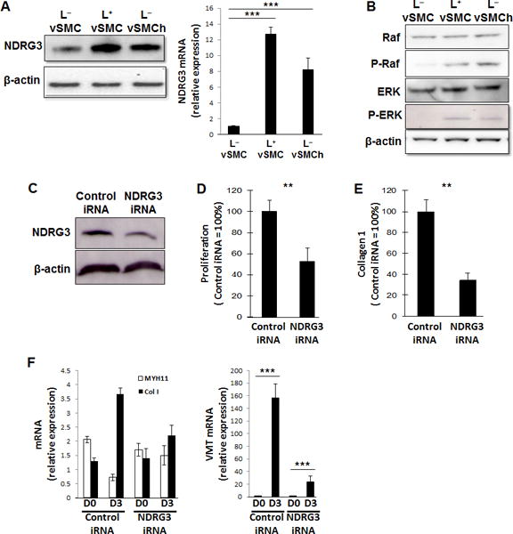
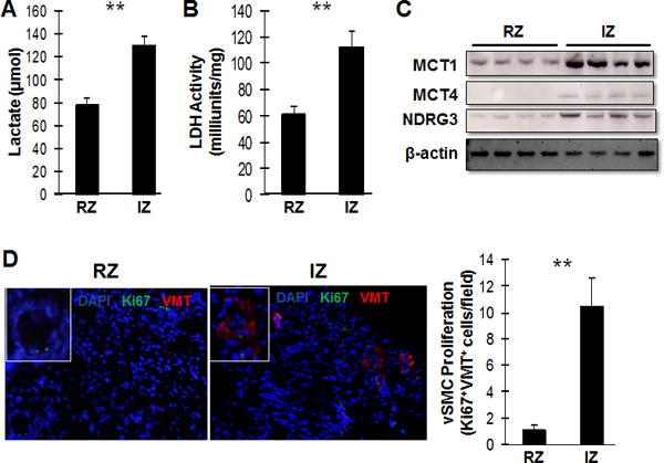
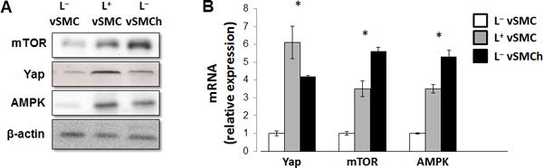
Methods and results: Experiments were performed with vSMCs that had been differentiated from human induced pluripotent stem cells and then cultured in glucose-free, lactate-enriched (L+) medium or in standard (L-) medium. Compared with the L- medium, the L+ medium was associated with significant increases in synthetic vSMC marker expression, proliferation, and migration and with significant declines in contractile and apoptotic activity. Furthermore, these changes were accompanied by increases in the expression of monocarboxylic acid transporters and were generally attenuated both by the blockade of monocarboxylic acid transporter activity and by transfection with iRNA for NDRG (N-myc downstream regulated gene). Proteomics, biomarker, and pathway analyses suggested that the L+ medium tended to upregulate the expression of synthetic vSMC markers, the production of extracellular proteins that participate in tissue construction or repair, and the activity of pathways that regulate cell proliferation and migration. Observations in hypoxia-cultured vSMCs were similar to those in L+-cultured vSMCs, and assessments in a swine myocardial infarction model suggested that measurements of lactate levels, lactate-dehydrogenase levels, vSMC proliferation, and monocarboxylic acid transporter and NDRG expression were greater in the ischemic zone than in nonischemic tissues.

Conclusions: These results demonstrate for the first time that vSMCs assume a more synthetic phenotype in a microenvironment that is rich in lactate. Thus, mechanisms that link glucose metabolism to vSMC phenotypic switching could play a role in the pathogenesis and treatment of cardiovascular disease.

Keywords: induced pluripotent stem cells; monocarboxylic acid transporters; myocardial infarction; phenotype; swine.


Figures

Figure 1
Figure 1. Lactate enhances hiPSC-vSMC proliferation
(A) Human aortic SMCs (HA-SMCs) and hiPSC-derived vSMCs (hiPSC-vSMCs) and ECs (hiPSC-ECs) were cultured in standard (i.e., lactate-free [L]) medium; then, intracellular glucose concentrations were determined and normalized to measurements in hiPSC-ECs. (B) HA-SMCs and hiPSC-vSMCs were cultured in L medium containing 5.5 mM or 22 mM glucose for 24 hours; then, the amount of glucose consumed and lactate produced was measured. (C) HA-SMCs, hiPSC-vSMCs, and hiPSC-ECs were cultured in glucose-free, lactate-containing (L+) medium for 20 minutes; then, intracellular lactate concentrations were determined and normalized to measurements in hiPSC-ECs. (D) hiPSC-SMCs and -ECs were cultured in L+ medium; then, intracellular ATP levels were measured at the indicated time points, normalized to the level observed at 0 h, and expressed as a percentage. (E) hiPSC-vSMCs and -ECs were cultured in L+ medium under normoxic (nmx) or hypoxic (hpx) conditions; then, intracellular lactate levels were determined at the indicated time points. (F) HA-SMCs and hiPSC-vSMCs were cultured in L+ or L medium for 4 days; live and dead cells were quantified at the beginning (D0) and end (D4) of the culture period and expressed as a percentage of the total number of cells at D0. (G) vSMCs were cultured with the indicated concentrations of lactate. Live and dead cells were quantified at the indicated time points and expressed as a percentage of the number observed at 0 h. *P<0.05, ** P<0.01.
Figure 2
Figure 2. Lactate promotes the synthetic phenotype in cultured hiPSC-vSMCs
(A) hiPSC-derived vSMCs (vSMCs) and human aortic smooth-muscle cells (HA-SMC) were cultured in L+ medium for 3 days, and their morphologies were evaluated at the beginning (D0) and end (D3) of the culture period via light microscopy; bar=100 μm (B) vSMCs were cultured in L+ medium (4 mM lactate) for three or four days; the expression of markers for the contractile (myosin heavy chain 11 [MYH11] and calponin [CNN]) and synthetic (collagen I [Col 1] and vimentin [VMT]) vSMC phenotype was evaluated via (i) quantitative RT-PCR (n=4) and (ii) Western blot at the beginning (D0) and end (D3 or D4) of the culture period. (C) vSMCs were cultured for 12 hours in standard (L) or L+ medium (i) with or (ii) without 3 ng/mL transforming growth-factor β; then, proliferation was evaluated via (i) optical density measurements of serially diluted cell solutions and (ii) immunofluorescent analyses of Ki67 expression; bar=20 μm. (D) vSMCs were cultured in L+ or L medium and then added with 300 uL of serum free medium to the upper chamber of a cell-migration apparatus. The lower chamber contained 500 μL medium and was coated with Matrigel, and the cells that had migrated into the lower chamber were quantified 12 hours later (n=3). (E) L+ and LvSMCs were plated onto collagen gels in 24-well culture dishes and allowed to attach overnight; then, the gels were released from the wells and images were obtained 0 and 24 hours later. Measurements at 24 hours were normalized to measurements at 0 hours and expressed as a percentage (n=3). (F) Apoptosis was evaluated in L+ and LvSMCs by quantifying caspase 3 expression, Bcl-2 expression, and Annexin V binding via (i) quantitative RT-PCR (n=3) and/or (ii) Western blot. (G) vSMCs were cultured in L+ or L medium for 24 hours and lysed; then, collagen levels were determined via ELISA (n=3). (H) vSMCs were cultured in L+ medium, in L medium, or in L medium under hypoxic conditions (vSMCh); then, the expression of (i) extracellular matrix proteins and (ii) transcription factors was evaluated via proteomics analysis. L+ medium vs L medium with hypoxia, **P<0.01, ***P<0.001 for all panels (n=3).
Figure 3
Figure 3. Lactate-induced phenotypic modulation of hiPSC-vSMCs is mediated by monocarboxylic acid transporters (MCTs)
(A) hiPSC-derived vSMCs were cultured in L+ medium (L+vSMC), in L medium (LvSMC), or in L medium under hypoxic conditions (LvSMCh); then, the expression of MCT1 and MCT4 were evaluated via Western blot (left) and quantitative RT-PCR (right). mRNA levels were normalized to measurements in LvSMC. (B-D) vSMCs were cultured in L+ medium, in L+ medium with the generalized MCT inhibitor α-CHC, or in L+ medium with the MCT1 inhibitor AZ3965 (AZ); then, (B) MCT1 and MCT4 expression was evaluated via Western blot (left) and quantitative RT-PCR (right), (C) proliferation was evaluated via proliferation assay (n= 3), and (D) collagen 1 levels were determined via ELISA (n= 3). mRNA levels were normalized to measurements in L+vSMC, and proliferation and collagen protein measurements were expressed as a percentage of measurements in cells that had been cultured in the absence of either inhibitor. (E) vSMCs were transfected with MCT4 iRNA or a scrambled iRNA sequence (Control iRNA) and cultured in L+ medium; then (i) MCT4 and MCT1 protein levels were evaluated via Western blot, (ii) proliferation was evaluated via proliferation assay (n= 3), and (iii) collagen 1 protein levels were evaluated via ELISA (n= 3); proliferation and collagen protein measurements were expressed as a percentage of measurements in cells that had been transfected with scrambled iRNA. (iv) MYH11, collagen 1 (Col 1), and vimentin (VMT) mRNA levels were evaluated via quantitative RT-PCR before (D0) and after (D3) a 3-day culture period (n= 3). **P<0.01 for all panels.
Figure 4
Figure 4. Lactate-induced phenotypic modulation of hiPSC-vSMCs is mediated by NDRG3
(A–B) hiPSC-derived vSMCs were cultured in L+ medium, in L medium, or in L medium under hypoxic conditions (LvSMCh); then, (A) the levels of NDRG3 protein (left) and mRNA (right) were evaluated via Western blot and quantitative RT-PCR (n=3), respectively, and (B) protein levels of Raf, phosphorylated Raf (P-Raf), (ERK), and phosphorylated ERK (P-ERK) were evaluated via Western blot. (C-F) vSMCs were transfected with NDRG3 iRNA or scrambled (control) iRNA. (C) NDRG3 protein levels in NDRG3-iRNA–transfected and control-iRNA–transfected vSMCs were evaluated via Western blot. (D-E) NDRG3-iRNA–transfected and control-iRNA–transfected SMCs were cultured in L+ medium; then, (D) proliferation was evaluated via proliferation assay (n=3) and (E) collagen 1 production was measured via ELISA (n=3). (F) NDRG3-iRNA–transfected and control-iRNA–transfected vSMCs were cultured in L+ medium for 3 days; mRNA levels of MYH11, collagen 1 (Col 1), and vimentin (VMT) were measured via quantitative RT-PCR before and after the culture period (n=3). **P<0.01, ***P<0.001 for all panels.
Figure 5
Figure 5. Lactate promotes the synthetic vSMC phenotype in the hearts of swine after MI
Myocardial infarction was surgically induced in the hearts of swine by occluding the left-anterior descending coronary artery for 60 minutes. (A) Lactate levels and (B) lactate dehydrogenase (LDH) levels were measured in the zone of ischemia (IZ) and in the remote (i.e., non-infarcted) zone (RZ) (n= 5). (C) MCT1, MCT4, and NDRG3 protein levels were evaluated in the IZ and RZ via Western blot. (D) Sections from the IZ and RZ were immunofluorescently stained for the presence of Ki67 (green) and vimentin (VMT, red), and nuclei were counterstained with DAPI (blue); then, the proliferation of vSMCs was evaluated by quantifying the number of cells that expressed both Ki67 and vimentin (n=5 sections per heart, 30 fields per section). **P<0.01 for all panels.
Figure 6
Figure 6. Lactate upregulates the expression of proteins involved in fibrosis and/or injury repair in vSMCs
hiPSC-derived vSMCs were cultured in L+ medium, in L medium, or in L medium under hypoxic conditions (LvSMCh); then, (A) the levels of mTOR, Yap, and AMPK (A) protein and (B) mRNA were evaluated via Western blot and quantitative RT-PCR, respectively (n=4). L mediumvs L+ medium and L medium with hypoxia all P<0.01 in RT-PCR, **P<0.01 for all panels.

References

    1. Owens GK, Kumar MS, Wamhoff BR. Molecular regulation of vascular smooth muscle cell differentiation in development and disease. Physiol Rev. 2004;84:767–801. - PubMed
    1. Yang L, Geng Z, Nickel T, Johnson C, Gao L, Dutton J, Hou C, Zhang J. Differentiation of human induced-pluripotent stem cells into smooth-muscle cells: Two novel protocols. PloS one. 2016;11:e0147155. - PMC - PubMed
    1. Bennett MR, Sinha S, Owens GK. Vascular smooth muscle cells in atherosclerosis. Circ Res. 2016;118:692–702. - PMC - PubMed
    1. Shi N, Chen SY. Smooth muscle cell differentiation: Model systems, regulatory mechanisms, and vascular diseases. J Cell Physiol. 2016;231:777–787. - PubMed
    1. Barron JT, Parrillo JE. Production of lactic acid and energy metabolism in vascular smooth muscle: Effect of dichloroacetate. Am J Physiol. 1995;268:H713–719. - PubMed

MeSH terms