Review
. 2017 Oct 11;37(41):9808-9818.
doi: 10.1523/JNEUROSCI.1788-16.2017.

Prying into the Prion Hypothesis for Parkinson's Disease

Affiliations
Review

Prying into the Prion Hypothesis for Parkinson's Disease

Patrik Brundin et al. J Neurosci. .

Abstract

In Parkinson's disease, intracellular α-synuclein inclusions form in neurons. We suggest that prion-like behavior of α-synuclein is a key component in Parkinson's disease pathogenesis. Although multiple molecular changes are involved in the triggering of the disease process, we propose that neuron-to-neuron transfer is a crucial event that is essential for Lewy pathology to spread from one brain region to another. In this review, we describe key findings in human postmortem brains, cultured cells, and animal models of disease that support the idea that α-synuclein can act as a prion. We consider potential triggers of the α-synuclein misfolding and why the aggregates escape cellular degradation under disease conditions. We also discuss whether different strains of α-synuclein fibrils can underlie differences in cellular and regional distribution of aggregates in different synucleinopathies. Our conclusion is that α-synuclein probably acts as a prion in human diseases, and a deeper understanding of this step in the pathogenesis of Parkinson's disease can facilitate the development of disease-modifying therapies in the future.Dual Perspectives Companion Paper: Parkinson's Disease Is Not Simply a Prion Disorder, by D. James Surmeier, José A. Obeso, and Glenda M. Halliday.

Keywords: Lewy body; alpha-synuclein; neurodegenerative disease; propagation; seeding.


Figures

Figure 1.
Figure 1.
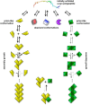
Mechanism of pathologic protein assembly and the notion of strains. Natively unfolded monomeric α-SYN multicolor molecule (top) populates different folding intermediates that expose specific amino acid stretches that determine their ability to establish defined sets of intermolecular interactions. This is schematized by the different shapes (folding intermediates) and colors (exposed amino acid stretches) of the folding intermediates (second row). The molecule in the conformation in yellow is capable of establishing longitudinal and lateral interactions with molecules in the same conformation. As long as only longitudinal or lateral interactions are established, the assemblies are transient. This is illustrated schematically by arrows, of the same size, in two directions. When both longitudinal and lateral interactions are established, a thermodynamically stable assembly is generated. This assembly can grow indefinitely by incorporation of molecules in the same conformation. This is illustrated schematically by a solid arrow pointing toward growth. Molecules constituting such an assembly can dissociate only from its ends; this is represented by a dashed arrow pointing toward disassembly. The molecule in the green conformation behaves in a similar manner, yielding fibrils made of green diamonds. The other conformers (red, blue, and purple) cannot establish thermodynamically stable intermolecular interactions. They do not yield assemblies. These folds can be considered as dead-end conformations. The intermolecular interactions the conformation in green is capable of establishing upon docking to an assembly made of yellow conformers (bottom), and vice versa, are unstable as they do not outweigh the entropic cost of binding. Thus, no mixed assemblies made of yellow and green conformers can form. The yellow and green assemblies expose distinct amino acid stretches that define among other things their interactomes, resistance to cellular clearance, tropism for different cell types in the nervous system, and the pathology they cause. The intermolecular interactions within the yellow and green assemblies and their surfaces that define their interactomes are at the origin of the notion of strains.
Figure 2.
Figure 2.
Schematic diagram depicting a possible central cascade leading to cell-to-cell transfer of α-SYN. The central process is likely affected by several other disease mechanisms that have already been implicated in PD (shown inside the circle) and that form an interdependent network of molecular events, which in combination or each on their own can promote the central cascade. Weak genetic risk factors and aging are depicted as potential triggers or promoters of cell-to-cell transfer of α-SYN (for details of the different mechanisms, see text).

References

    1. Abounit S, Bousset L, Loria F, Zhu S, de Chaumont F, Pieri L, Olivo-Marin JC, Melki R, Zurzolo C (2016) Tunneling nanotubes spread fibrillar α-synuclein by intercellular trafficking of lysosomes. EMBO J 35:2120–2138. 10.15252/embj.201593411 - DOI - PMC - PubMed
    1. Allen Reish HE, Standaert DG (2015) Role of α-synuclein in inducing innate and adaptive immunity in Parkinson disease. J Parkinsons Dis 5:1–19. 10.3233/JPD-140491 - DOI - PMC - PubMed
    1. Alvarez-Erviti L, Seow Y, Schapira AH, Gardiner C, Sargent IL, Wood MJ, Cooper JM (2011) Lysosomal dysfunction increases exosome-mediated alpha-synuclein release and transmission. Neurobiol Dis 42:360–367. 10.1016/j.nbd.2011.01.029 - DOI - PMC - PubMed
    1. Angot E, Steiner JA, Hansen C, Li JY, Brundin P (2010) Are synucleinopathies prion-like disorders? Lancet Neurol 9:1128–1138. 10.1016/S1474-4422(10)70213-1 - DOI - PubMed
    1. Angot E, Steiner JA, Lema Tomé CM, Ekström P, Mattsson B, Björklund A, Brundin P (2012) Alpha-synuclein cell-to-cell transfer and seeding in grafted dopaminergic neurons in vivo. PLoS One 7:e39465. 10.1371/journal.pone.0039465 - DOI - PMC - PubMed

Publication types