. 2017 Dec 1;28(25):3647-3659.
doi: 10.1091/mbc.E17-08-0497. Epub 2017 Oct 11.

A microtubule polymerase cooperates with the kinesin-6 motor and a microtubule cross-linker to promote bipolar spindle assembly in the absence of kinesin-5 and kinesin-14 in fission yeast

Affiliations

A microtubule polymerase cooperates with the kinesin-6 motor and a microtubule cross-linker to promote bipolar spindle assembly in the absence of kinesin-5 and kinesin-14 in fission yeast

Masashi Yukawa et al. Mol Biol Cell. .

Abstract

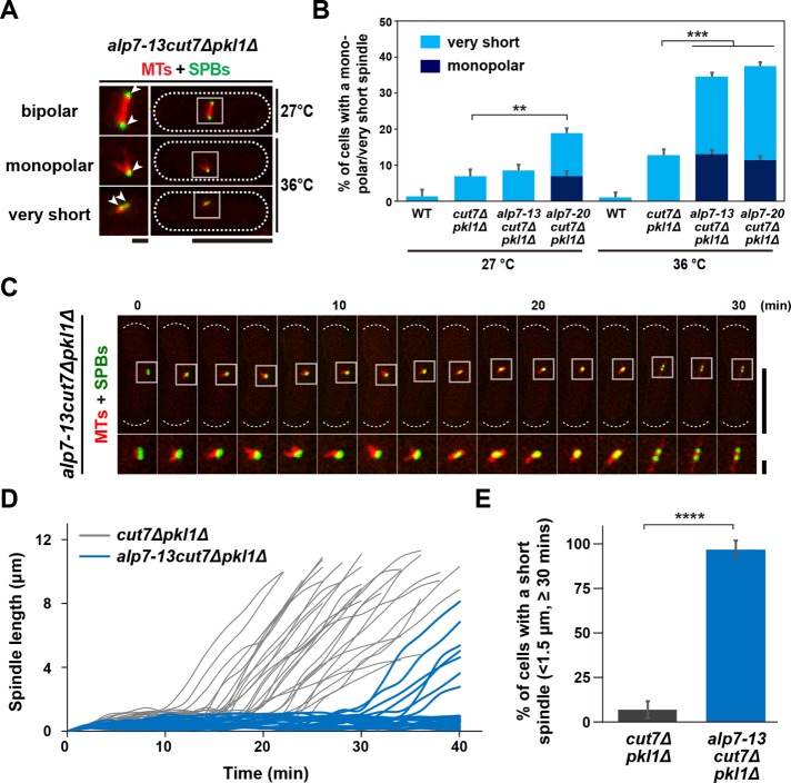
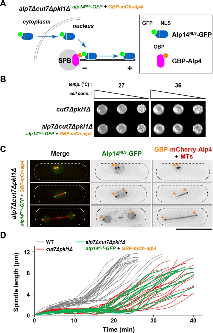
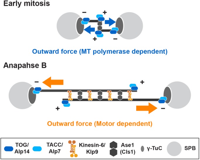
Accurate chromosome segregation relies on the bipolar mitotic spindle. In many eukaryotes, spindle formation is driven by the plus-end-directed motor kinesin-5 that generates outward force to establish spindle bipolarity. Its inhibition leads to the emergence of monopolar spindles with mitotic arrest. Intriguingly, simultaneous inactivation of the minus-end-directed motor kinesin-14 restores spindle bipolarity in many systems. Here we show that in fission yeast, three independent pathways contribute to spindle bipolarity in the absence of kinesin-5/Cut7 and kinesin-14/Pkl1. One is kinesin-6/Klp9 that engages with spindle elongation once short bipolar spindles assemble. Klp9 also ensures the medial positioning of anaphase spindles to prevent unequal chromosome segregation. Another is the Alp7/TACC-Alp14/TOG microtubule polymerase complex. Temperature-sensitive alp7cut7pkl1 mutants are arrested with either monopolar or very short spindles. Forced targeting of Alp14 to the spindle pole body is sufficient to render alp7cut7pkl1 triply deleted cells viable and promote spindle assembly, indicating that Alp14-mediated microtubule polymerization from the nuclear face of the spindle pole body could generate outward force in place of Cut7 during early mitosis. The third pathway involves the Ase1/PRC1 microtubule cross-linker that stabilizes antiparallel microtubules. Our study, therefore, unveils multifaceted interplay among kinesin-dependent and -independent pathways leading to mitotic bipolar spindle assembly.


Figures

FIGURE 1:
FIGURE 1:
The cut7pkl1 double deletion results in SAC-dependent mitotic delay with a short preanaphase spindle. (A) Indicated strains were streaked on a rich YE5S agar plate containing Phloxine B and incubated at 33°C for 2 d. (B) Kymographic images of mitotic wild-type (top) and cut7∆pkl1∆ cells (bottom) expressing a tubulin marker (mCherry-Atb2, red) and an SPB marker (Cut12-GFP, green). Pictures were taken at 2 min intervals. Scale bar, 10 μm. (C) Profiles of mitotic progression in wild-type (gray line, n = 27) and cut7∆pkl1∆ cells (red line, n = 24). Changes of the inter-SPB distance were plotted against time. (D) The time between the initiation of SPB separation and onset of anaphase B. (E) Spindle length at anaphase onset. (F) Spindle growth rate during anaphase B. Results are given as means ± SD. Data sets were compared with a two-tailed unpaired Student’s t tests (****p < 0.0001). (G) Tetrad analysis on crosses between mad1∆pkl1∆ and cut7∆pkl1∆ strains, Individual spores (ad) in each ascus (14) were dissected on YE5S plates and incubated at 27°C for 3 d. Representative tetrad patterns are shown. Circles, triangles and squares with green lines indicate pkl1∆ single mutants, cut7∆pkl1∆ double mutants and mad1∆pkl1∆ double mutants, respectively. Assuming 2:2 segregation of individual markers allows the identification of mad1∆cut7∆pkl1∆ triple mutants (indicated by dashed magenta circles).
FIGURE 2:
FIGURE 2:
Motor activity of Kinesin-6/Klp9 is essential for the viability in the absence of Cut7 and Pkl1. (A, B, C) Tetrad analysis. Spores were dissected on crosses between klp9∆pkl1∆ and cut7∆pkl1∆ strains (A), between klp9rigorpkl1∆ and cut7∆pkl1∆ strains (B), or between klp9∆38Cpkl1∆ and cut7∆pkl1∆ strains (C) respectively. Manipulation was performed as in Figure 1G. Circles and triangles with green lines indicate pkl1∆ single mutants and cut7∆pkl1∆ double mutants, respectively. Squares with green lines indicate klp9∆pkl1∆ double mutants (A), klp9rigorpkl1∆ double mutants (B), or klp938Cpkl1∆ double mutants (C). Assuming 2:2 segregation of individual markers allows the identification of triple mutants in each cross (indicated by dashed magenta circles). (D) Kymographic images of a wild-type, pkl1∆, or cut7∆pkl1∆ cell expressing Klp9-GFP (green) along with a kinetochore marker (Mis6-2mRFP, red) (Saitoh et al., 1997) and an SPB marker (Pcp1-CFP, blue) (Flory et al., 2002; Fong et al., 2010). Images were taken at 2-min intervals. The timing of SPB separation, anaphase onset, and Klp9-GFP accumulation at the spindle midzone is indicated with white, magenta, and yellow arrowheads, respectively. Scale bar, 10 μm. (E) Time-lapse images of a mitotic cut7-22pkl1∆klp9∆ cell. Spindle microtubules (mCherry-Atb2; red) and SPB (Cut12-GFP; green) were visualized (top). Images were taken at 2-min intervals on shifting the temperature from 27°C to 36°C for 2 h. The septation sites are indicated with yellow arrowheads in the DIC images (bottom). The cell peripheries are outlined with dotted lines (top). Scale bar, 10 μm. (F, G) Profiles of mitotic progression in cut7∆pkl1∆ (gray, n = 30) and cut7-22pkl1∆klp9∆ cells (yellow, n = 21) under the same condition as in E. Changes of the inter-SPB distance were plotted against time (F). Spindle growth rate after the inter-SPB distance reached 1.5 μm was calculated (G). Data are given as means ± SD; ****p < 0.0001 (two-tailed unpaired Student’s t test).
FIGURE 3:
FIGURE 3:
The point mutations within the Klp9 motor domain interfere with elongation and positioning of anaphase B spindle in cut7∆pkl1∆. (A) Mutation sites in the klp9 mutants. Klp9-3 contained two separate mutations. Then two strains containing one of them were created (Klp9-3M and Klp9-3T). (B) Spot test. Indicated strains were serially (10-fold) diluted, spotted onto rich YE5S plates, and incubated at 27°C or 36°C for 2 d. cell conc., cell concentration; temp., temperature. (C) Synchronous culture analysis with centrifugal elutriation. Small G2 cells of cut7∆pkl1∆ or klp9-2cut7∆pkl1∆ grown at 27°C were collected with centrifugal elutriation and shifted to 36°C at time 0. Spindle microtubules (mCherry-Atb2) and SPB (Cut12-GFP) were visualized to analyze mitotic progression. The percentages of mitotic cells (red line) and cells with the septum (green line) were counted to monitor cell cycle progression. Relative colony-forming units at the indicated time points were assessed as cell viability (=100% at time 0; gray line). Aliquots of cell cultures at each time point were stained with Hoechst33342 to detect chromosome segregation defects (light blue line). Light red columns mark periods in mitosis. (D) Time-lapse images of mitotic klp9-2cut7∆pkl1∆ cells. Spindle microtubules (mCherry-Atb2; red) and SPB (Cut12-GFP; green) were visualized. Images were taken at 2 min intervals after incubation of cultures at 36°C for 6 h. The septation sites are indicated with yellow arrowheads. The cell peripheries are outlined with dotted lines (top). Scale bar, 10 μm. (E, F) Profiles of mitotic progression in klp9-2cut7∆pkl1∆ (orange, n = 34) cells under the same condition as in D. Changes of the inter-SPB distance are plotted against time (E). Spindle growth rate after the inter-SPB distance reached 1.5 μm was calculated (F). Data for cut7∆pkl1∆ is shown for comparison; this is the same as that presented in Figure 2, F and G. Data are given as means ± SD; ****p < 0.0001 (two-tailed unpaired Student’s t test).
FIGURE 4:
FIGURE 4:
SPB localization of Alp7/TACC is essential for proper spindle assembly in the cut7∆pkl1∆ mutant. (A) List of MAPs tested for synthetic lethality (SL) when combined with cut7∆pkl1∆. Information of each protein function and synthetic lethality is given. (B) Truncations or point mutations of Alp7 tested for synthetic lethality with cut7∆pkl1∆. Information of SPB localization and synthetic lethality is given. See Supplemental Figure S5A for more details. (C) Spot test. Indicated strains were serially (10-fold) diluted, spotted onto rich YE5S plates and incubated at 27°C, 33°C, or 36°C for 2 d. cell conc., cell concentration; temp., temperature. (D) Intensity of Alp7-GFP localized to the mitotic SPB in the alp7 mutants. Representative images showing mitotic localization of Alp7-GFP in the indicated strains are presented at the top. Insets 1 and 2 show the magnified regions of the SPB. Cells of wild type (carrying Alp7-GFP and mCherry-Atb2) and alp7-13 or alp7-20 (carrying Alp7-GFP and Pcp1-2mRFP) were mixed in the same culture, shifted from 27°C to 36°C, and incubated for 2 h (see Supplemental Figure S5C). Quantification of signal intensities of Alp7-GFP at the SPB is shown at the bottom. All p values were obtained from the two-tailed unpaired Student’s t test. Data are presented as the means ± SD (≥20 cells). ****p < 0.0001. The cell peripheries are outlined with dotted lines. Scale bars, 10 μm (main images) and 1 μm (enlarged images of the boxed regions on the right). a.u., arbitrary unit.
FIGURE 5:
FIGURE 5:
Alp7 is required for bipolar spindle assembly during early mitosis of the cut7∆pkl1∆ cells. (A) Representative spindle morphologies of the alp7-13cut7∆pkl1∆ triple mutants. Cells containing a bipolar (top), a monopolar (middle), or a very short spindle (the inter-SPB distance is <0.5 μm, bottom) are shown (Cut12-GFP, green, and mCherry-Atb2, red). Enlarged images of spindle areas (squares) are shown on the left panels. Positions of the SPB are indicated with arrowheads. The cell peripheries are outlined with dotted lines. Scale bars, 10 μm (main images) and 1 μm (enlarged images of the boxed regions). (B) The percentage of cells containing a monopolar spindle (dark blue) or a very short spindle (light blue). Each strain was grown at 27°C and shifted to 36°C for 2 h. In each experiment, more than 25 mitotic cells were observed. (C) Time-lapse images of a mitotic alp7-13cut7∆pkl1∆ cell. Spindle microtubules (mCherry-Atb2; red) and SPBs (Cut12-GFP; green) were visualized. Images were taken at 2-min intervals after 2 h incubation at 36°C. The positions of cell ends are indicated with dotted curved lines (top), and enlarged images of the boxed regions are presented at the bottom. Scale bars, 10 μm (top) and 1 μm (bottom). (D, E) Profiles of mitotic progression in alp7-13cut7∆pkl1∆ (light blue, n = 26) cells under the same condition as in C. Changes in the inter-SPB distance were plotted against time (D). The percentage of cells retaining a short spindle (the inter-SPB distance is <1.5 μm) for >30 min after SPB separation was counted (E). Data for cut7∆pkl1∆ are shown for comparison; this is the same as that presented in Figure 2, F and G. Data are given as means ± SD; **p < 0.01; ***p < 0.001; ****p < 0.0001 (two-tailed unpaired Student’s t test).
FIGURE 6:
FIGURE 6:
Tethering of Alp14/TOG to mitotic SPB in the alp7∆cut7∆pkl1∆ cells enables Cut7-independent bipolar spindle formation. (A) Schematic illustration of a strategy for the forced tethering of Alp14 to the SPB in the absence of Alp7. In wild-type cells, Alp7 forms a complex with Alp14 in the cytoplasm and imports Alp14 into the nucleus and recruits it to the mitotic SPB. In the absence of Alp7, Alp14 cannot enter the nucleus nor does it localize to the SPB. However, even without Alp7, GFP- and NLS-tagged Alp14 (Alp14NLS-GFP) could enter the nucleus, where it would be tethered to the SPB through the interaction with GBP-tagged Alp4, a core component of the γ-tubulin complex (Vardy and Toda, 2000). (B) Rescue of otherwise lethal alp7∆cut7∆pkl1∆ triple mutants by forced tethering of Alp14 to the SPB. Serial dilution spot tests were performed using the indicated strains on rich YE5S media, and the cells were incubated at 27°C or 36°C for 2 d. cell conc., cell concentration; temp., temperature. (C) Visualization of Alp14NLS-GFP localization. Representative images of mitotic alp7∆cut7∆pkl1∆ cells containing Alp14NLS-GFP (green), GBP-mCherry-Alp4 (red) and mCherry-Atb2 (red) are shown. The positions of SPBs are indicated with orange arrowheads. The cell peripheries are outlined with dotted lines. Some Alp14NLS-GFP signals are detected in the nucleus and along the microtubules in addition to SPBs. Scale bar, 10 μm. (D) Profiles of mitotic progression in alp7∆cut7∆pkl1∆ cells containing SPB-tethered Alp14NLS-GFP. Changes of the inter-SPB distance are plotted against time (green lines; n = 14). Data for wild-type (thin gray lines) and cut7∆pkl1∆ (thin red lines) are the same as those shown in Figure 1C. See Supplemental Figure S7, A–C, for quantitative data.
FIGURE 7:
FIGURE 7:
Model of bipolar spindle formation in the absence of kinesin-5/Cut7 and kinesin-14/Pkl1. In the absence of Cut7Kin-5 and Pkl1Kin-14, three distinct pathways compensate for defective spindle assembly and lethality in fission yeast. In early mitosis (top), the Alp7/TACC-Alp14/TOG microtubule polymerase complex is localized to the SPB, by which Alp14 elongates spindle microtubules. Alp14 may also promote microtubule nucleation reaction from the SPB (Reber et al., 2013; Roostalu et al., 2015; Hussmann et al., 2016). Interaction of growing microtubule plus ends with the other SPB would generate outward force in place of Cut7Kin-5. This force is sufficient to separate duplicated SPBs, thereby promoting short bipolar spindle formation in the absence of Pkl1/kinesin-14-dependent inward force. On onset of anaphase B (bottom), Klp9Kin-6 is localized to the overlapping zones within short spindles and increases spindle length by sliding them apart toward both cell ends. For simplicity, the requirement for this kinesin-6 for CPC recruitment to the spindle midzone is not depicted in this figure. Klp9Kin-6 is also important for the medial positioning of elongating spindles. Ase1/PRC1 is localized to the spindle microtubules and cross-links antiparallel microtubules to ensure the establishment of bipolarity. Cls1/CLASP may function in stabilization of bipolar spindles in cooperation with Ase1 (Bratman and Chang, 2007; Rincon et al., 2017).

Similar articles

Cited by

References

    1. Abaza A, Soleilhac JM, Westendorf J, Piel M, Crevel I, Roux A, Pirollet F. M phase phosphoprotein 1 is a human plus-end-directed kinesin-related protein required for cytokinesis. J Biol Chem. 2003;278:27844–27852. - PMC - PubMed
    1. Adams RR, Tavares AA, Salzberg A, Bellen HJ, Glover DM. pavarotti encodes a kinesin-like protein required to organize the central spindle and contractile ring for cytokinesis. Genes Dev. 1998;12:1483–1494. - PMC - PubMed
    1. Al-Bassam J, Chang F. Regulation of microtubule dynamics by TOG-domain proteins XMAP215/Dis1 and CLASP. Trends Cell Biol. 2011;21:604–614. - PMC - PubMed
    1. Al-Bassam J, Kim H, Brouhard G, van Oijen A, Harrison SC, Chang F. CLASP promotes microtubule rescue by recruiting tubulin dimers to the microtubule. Dev Cell. 2010;19:245–258. - PMC - PubMed
    1. Al-Bassam J, Kim H, Flor-Parra I, Lal N, Velji H, Chang F. Fission yeast Alp14 is a dose dependent plus end tracking microtubule polymerase. Mol Biol Cell. 2012;23:2878–2890. - PMC - PubMed

MeSH terms

LinkOut - more resources