. 2017 Oct 11;7(1):12953.
doi: 10.1038/s41598-017-13072-1.

Danshenol A inhibits TNF-α-induced expression of intercellular adhesion molecule-1 (ICAM-1) mediated by NOX4 in endothelial cells

Affiliations

Danshenol A inhibits TNF-α-induced expression of intercellular adhesion molecule-1 (ICAM-1) mediated by NOX4 in endothelial cells

Wenwen Zhao et al. Sci Rep. .

Abstract

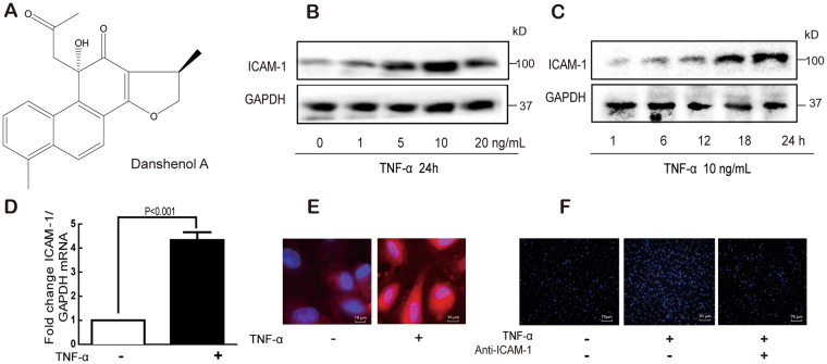
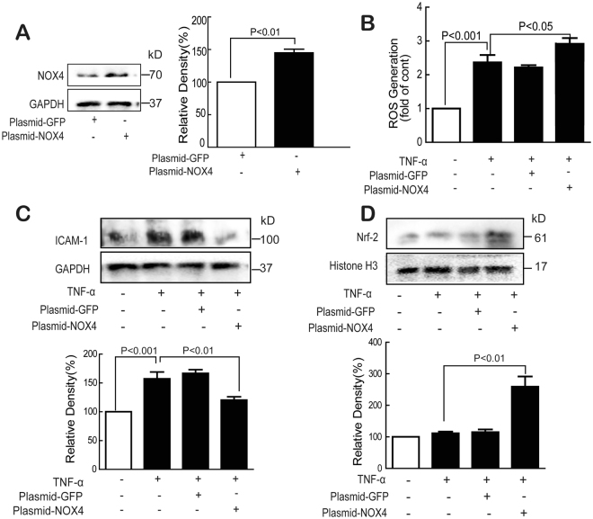
ICAM-1 overexpression and subsequent adhesion of leukocytes to endothelial cells play critical roles in the early stage of atherosclerosis. Danshenol A (DA) is an abietane-type diterpenoid isolated from traditional Chinese herb Salvia miltiorrhiza Bunge. The mechanisms under its regulation of adhesion of molecular expression are explored. Here, the effect of DA on TNF-α-induced ICAM-1 expression was investigated in endothelial cells. TNF-α-induced ICAM-1 expression and subsequent adhesion of monocytes, as well as elevated reactive oxygen species (ROS) generation and NOX4 expression were all significantly reversed by DA, siNOX4 and NOX4 inhibitor GKT137831. Furthermore, TNF-α-induced ICAM-1 expression, which was increased via IKKβ/IκBα-mediated activation of NF-κB p65, was also inhibited by DA. Interestingly, NOX4 overexpression suppressed the ICAM-1 expression, and this finding may be ascribed to the activation of Nrf-2. Additionally, NF-κB inhibitor PDTC, siNOX4, or DA can decrease the TNF-α-induced ICAM-1 expression and suppress the adhesion of monocytes. In all, DA inhibited TNF-α-induced ICAM-1 expression and subsequent monocyte adhesion to endothelial cells through the NOX4-dependent IKKβ/NF-κB pathway. Besides, NOX4 played dual role in regulating ICAM-1 expression via diverse signal pathway. This novel bioactivity will make DA a good candidate to be further explored for therapeutic or preventive application for atherosclerosis.


Conflict of interest statement

The authors declare that they have no competing interests.

Figures

Figure 1
Figure 1
TNF-α induces ICAM-1 expression and monocyte–HUVEC adhesion. Chemical structure of DA (A). Cells were treated with TNF-α, and the ICAM-1 protein expression was detected by Western blot (B and C). Cells were treated with TNF-α (10 ng/mL) for 24 h; ICAM-1 mRNA and protein expression were detected by real-time PCR (D) and immunofluorescence staining (E), respectively. Cells were treated with TNF-α (10 ng/mL) for 24 h, and the adhesion of monocytes to endothelial cells was determined with or without ICAM-1 antibody (F). Data were presented as mean ± SD of three independent experiments.
Figure 2
Figure 2
DA inhibited TNF-α-induced ICAM-1 expression and monocyte–HUVEC adhesion. Cells were treated with DA, and cytotoxicity was determined by MTT assay (A). Cells were treated with DA (10 nM) for 24 h; ICAM-1 expression was detected by Western blot (B) and RT-PCR (C). Cells were treated with TNF-α (10 ng/mL) for 24 h in the absence or presence of DA (10 nM) pretreatment for 1 h; ICAM-1 protein and mRNA expression was detected by Western blot (D), immunofluorescence staining (F), and RT-PCR (E). The effect of DA on the adhesion of monocytes to endothelial cells was determined (G). Data were presented as mean ± SD of three independent experiments. NS indicates no significance.
Figure 3
Figure 3
DA restored TNF-α-induced endothelial redox imbalance by inhibiting TNF-α-induced NADPH oxidase activity. Cells were treated with TNF-α (10 ng/mL) for 1 h with or without DA (10 nM), NAC (5 mM), Rot (20 μM), TTFA (10 μM), AA (5 μM), DPI (1 μM), All (10 μM), or NDGA (10 μM) pretreatment for 1 h, and the generation of ROS, H2O2, and O2 •− was determined by DCFH2-DA (A and F), Amplex Red (B), and DHE (C), respectively. GSH/GSSG levels were measured using commercial kits (D). DPPH radical scavenging activity was determined in a cell-free system (E). Data were presented as mean ± SD of three independent experiments.
Figure 4
Figure 4
DA inhibited TNF-α-induced NADPH oxidase expression. Cells were treated with TNF-α (10 ng/mL) for 24 h with or without DA (10 nM) pretreatment for 1 h, and the protein expression of NOX1, NOX2, NOX4, and p22phox was detected by Western blot (A,B,D and E) or immunofluorescence (C). Data were presented as mean ± SD of three independent experiments. NS indicates no significance.
Figure 5
Figure 5
Knockdown of NOX4 inhibited TNF-α-induced ICAM-1 expression in endothelial cells. NOX4 was silenced by siNOX4 or inhibitor GKT137831 (A and B); After NOX4 was silenced, cells were treated with TNF-α (10 ng/mL) and ROS generation was detected by flow cytometry (C) and ICAM-1 was determined by Western blot (D); The constructed model of NOX4 (E); Ramachandran plot for NOX4 (F); Dark green dots represent the residues in most favored regions, yellow dots represent the residues in allowed regions; The ligand interaction of DA with NOX4 (G); The binding model of DA with NOX4 (K). The ligand is colored in blue, and the surrounding residues in the binding pockets are colored in orange. Data were presented as mean ± SD of three independent experiments.
Figure 6
Figure 6
DA inhibited NF-κB pathways in endothelial cells. Cells were treated with TNF-α (10 ng/mL) for 1 h with or without DA (10 nM) pretreatment; the expression of Nrf-1, Nrf-2, and NF-κB p65 in the nucleus was detected by Western blot (A) and immunofluorescence staining (B). The expression of p-IκBα, IκBα, p-IKKβ, and IKKβ was also determined by Western blot (C). The interaction of IκBα and IKKβ was determined by co-immunoprecipitation assay (D). Cells were treated with TNF-α (10 ng/mL) for 1 h in the absence or presence of NAC (5 mM) for 1 h; the expression of IKKβ was detected by Western blot (E). Data were presented as mean ± SD of three independent experiments. NS indicates no significance.
Figure 7
Figure 7
Overexpression of NOX4 inhibited TNF-α-induced ICAM-1 expression. Normal and NOX4 overexpressed cells (A) were treated with TNF-α (10 ng/mL) for 24 h, and the expression of NOX4, ICAM-1 and nuclear Nrf-2 (A,C and D), ROS generation (B) were determined by Western blot and flow cytometry. Data were presented as mean ± SD of three independent experiments.
Figure 8
Figure 8
DA inhibited monocyte adhesion by decreasing ICAM-1 expression through NOX4/NF-κB signaling. Cells were treated with TNF-α (10 ng/mL) for 24 h with or without DA (10 nM), siNOX4, or PDTC (10 μM) pretreatment for 1 h, and the ICAM-1 protein (A) and mRNA (B) expression were determined by Western blot and RT-PCR; The adhesion of monocytes to endothelial cells was performed (C). Data were presented as mean ± SD of three independent experiments.
Figure 9
Figure 9
Schematic diagram illustrating the molecular mechanisms underlying the anti-ICAM-1 of DA in endothelial cells.

References

    1. Tuttolomondo A, et al. Atherosclerosis as an Inflammatory Disease. Curr Pharm Design. 2012;18:4266–4288. doi: 10.2174/138161212802481237. - DOI - PubMed
    1. Chen YH, Lin SJ, Chen JW, Ku HH, Chen YL. Magnolol attenuates VCAM-1 expression in vitro in TNF-alpha-treated human aortic endothelial cells and in vivo in the aorta of cholesterol-fed rabbits. British journal of pharmacology. 2002;135:37–47. doi: 10.1038/sj.bjp.0704458. - DOI - PMC - PubMed
    1. Krunkosky TM, et al. Effects of TNF-alpha on expression of ICAM-1 in human airway epithelial cells in vitro. Signaling pathways controlling surface and gene expression. American journal of respiratory cell and molecular biology. 2000;22:685–692. doi: 10.1165/ajrcmb.22.6.3925. - DOI - PubMed
    1. Zhao, W., Wu, C. & Chen, X. Cryptotanshinone Inhibits oxidized LDL-Induced Adhesion Molecule Expression via ROS Dependent NF-kappaB Pathways. Cell adhesion & migration, 0, 10.1080/19336918.2015.1119361 (2015). - PMC - PubMed
    1. Osterud B, Bjorklid E. Role of monocytes in atherogenesis. Physiological reviews. 2003;83:1069–1112. doi: 10.1152/physrev.00005.2003. - DOI - PubMed

Publication types

MeSH terms