. 2017 Oct 11;8(1):853.
doi: 10.1038/s41467-017-00889-7.

The metabolic enzyme fructose-1,6-bisphosphate aldolase acts as a transcriptional regulator in pathogenic Francisella

Affiliations

The metabolic enzyme fructose-1,6-bisphosphate aldolase acts as a transcriptional regulator in pathogenic Francisella

Jason Ziveri et al. Nat Commun. .

Abstract

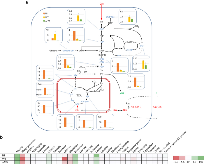
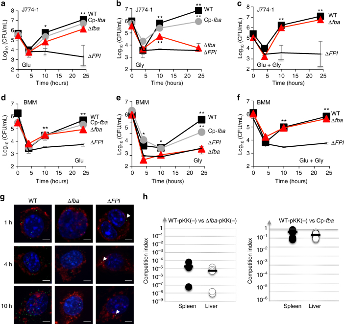
The enzyme fructose-bisphosphate aldolase occupies a central position in glycolysis and gluconeogenesis pathways. Beyond its housekeeping role in metabolism, fructose-bisphosphate aldolase has been involved in additional functions and is considered as a potential target for drug development against pathogenic bacteria. Here, we address the role of fructose-bisphosphate aldolase in the bacterial pathogen Francisella novicida. We demonstrate that fructose-bisphosphate aldolase is important for bacterial multiplication in macrophages in the presence of gluconeogenic substrates. In addition, we unravel a direct role of this metabolic enzyme in transcription regulation of genes katG and rpoA, encoding catalase and an RNA polymerase subunit, respectively. We propose a model in which fructose-bisphosphate aldolase participates in the control of host redox homeostasis and the inflammatory immune response.The enzyme fructose-bisphosphate aldolase (FBA) plays central roles in glycolysis and gluconeogenesis. Here, Ziveri et al. show that FBA of the pathogen Francisella novicida acts, in addition, as a transcriptional regulator and is important for bacterial multiplication in macrophages.


Conflict of interest statement

The authors declare no competing financial interests.

Figures

Fig. 1
Fig. 1
Carbon isotopologue distribution of central metabolites in BMMs. a Absolute concentrations of central metabolites in intracellular cell extracts (in µmol L−1) after 24 h of cultivation of BMM macrophages: NI (n = 1); or infected either with wild-type F. novicida (WT) (n = 3) or ∆FPI (∆FPI) strain (n = 3). Fumarate measurements (Fum) represent MS peak area instead of absolute concentrations. b Consumed and produced metabolites, measured in extracellular media in the different infection conditions, profiled by NMR. Mean concentration changes, presented here by substracting concentration measured at 24 h to those measured at 1 h. Absolute concentrations changes are expressed in mM. Positive/green values correspond to metabolites accumulated in the media, negative/red values correspond to metabolites consumed by cells from the media between 1 and 24 h of incubation. Analyses were performed on biological triplicates and each sample was run in technical triplicates (mean and SD of metabolite concentrations were calculated using R 3.2.3, R Foundation for Statistical Computing, Vienna, Austria. URL http://www.R-project.org/)
Fig. 2
Fig. 2
FBA inactivation inhibits growth of Francisella in the presence of various carbon sources. Wild-type F. novicida (WT, close black squares), isogenic ∆fba mutant (Δfba, red circles), and complemented fba strain (Δfba Cp-fba, gray line), and isogenic ∆glpX mutant (ΔglpX, blue diamond), were grown in chemically defined medium (CDM) lacking glucose and supplemented with different carbon source at a final concentration of 25 mM. a Glucose (Glc); b Pyruvate (Pyr); c Glycerol (Gly); d Ribose (Rib); e Fructose (Fru); f Succinate (Suc); g N-Acetylglucosamine (GlcNAc); h Glucose + Pyruvate (Glc + Pyr); i Glucose + Glycerol (Glc + Gly)
Fig. 3
Fig. 3
fba inactivation affects intracellular survival. Intracellular bacterial multiplication of wild-type F. novicida (WT, black squares), isogenic ∆fba mutant (ΔFBA, red triangles), and complemented Δfba strain (Cp-fba, gray circles), and the ΔFPI negative control (black lines), was monitored during 24 h in J774A.1 macrophage cells and bone marrow-derived macrophages (BMM) from 6 to 8-week-old female BALB/c mice. DMEM (Dulbecco’s modified eagle medium) was supplemented either with glucose a, d, glycerol b, e, or an equimolar concentration of glucose and glycerol c, f. af mean and SD of triplicate wells are shown. Each sugar was added to the cell culture medium at a final concentration of 5 mM. *p < 0.01; **p < 0.001 (determined by two-tailed unpaired Student’s t-test). g Glycerol-grown J774.1 were infected for 30 min with wild-type F. novicida (WT), Δfba, or ΔFPI strains and their co-localization with the phagosomal marker LAMP-1 was observed by confocal microscopy 1, 4, and 10 h, after beginning of the experiment. Scale bars at the bottom right of each panel correspond to 5 μM. J774.1 were stained for F. tularensis (green), LAMP-1 (red), and host DNA (blue, DAPI stained). h Group of five BALB/c mice were infected intraperitoneally with 100 CFU of wild-type F. novicida and 100 CFU of Δfba mutant strain. Bacterial burden was realized in liver (open circles, right column) and spleen (black circles, left column) of mice. The data represent the competitive index (CI) value (in ordinate) for CFU of mutant/wild-type of each mouse, after 48 h infection, divided by CFU of mutant/wild-type in the inoculum. Bars represent the geometric mean CI value
Fig. 4
Fig. 4
Oxidative stress survival. After an overnight culture in CDM supplemented with glucose and glycerol, bacteria were diluted in PBS and were subjected to oxidative stress a 10 mM H202, b 500 µM H202, c Tert-butyl hydroperoxide 10 mM (TBH), and d Cumene hydroperoxide 10 mM (CH). Bacteria were plated on chocolate agar plates at different time points and viable bacteria were numerated 2 days after. Experiments were realized three times
Fig. 5
Fig. 5
Quantitative real-time RT-PCR analysis. a, b Bacteria were grown overnight in TSB and qRT-PCR analyses were performed on selected genes, a katG gene or b fba gene in wild-type F. novicida, Δfba mutant, and fba-complemented (Cp-fba) strains. For each gene, the transcripts were normalized to helicase rates (FTN_1594). c Catalase activity assay was realized at 570 nm, using the Catalase Assay Kit (ab83464; Abcam). The assay was performed according to manufacturer’s recommendation. Each assay was performed on two independent protein lysates. The average of OD570 ± SD was recorded for wild-type F. novicida and the Δfba mutant. The catalase activity recorded in the ∆fba mutant was significantly lower than that recorded in the wild-type strain (**p < 0.01). d J774.1 cells were infected for 24 h with wild-type F. novicida (WT) and Δfba mutant. Total RNA was analyzed by quantitative RT-PCR with katG and fba gene. For each gene, the transcripts were normalized to helicase rates (FTN_1594) (**p < 0.01). e J774.1 cells were infected for 24 h with wild-type F. novicida (WT) and Δfba mutant in DMEM supplemented with glucose and glycerol (25 mM). The supernatants were analyzed by ELISA to detect the amounts of IL-6 produced at 24 h in pg mL−1. f Intracellular bacterial multiplication of wild-type F. novicida (WT) and ∆fba mutant was determined at 24 h, in the infected J774.1 cells used for the IL-6 dosage, as a control. (**p < 0.01). ac mean and SD of three independent experiments are shown; df each assay was repeated at least three times. Mean and SD of three wells of one typical experiment are shown. p-values were determined by the two-tailed unpaired Student’s t-test
Fig. 6
Fig. 6
Biclustering and heat map of differential proteins. Heat map and biclustering were obtained by comparing the 26 differentially expressed proteins identified from label-free MS analysis of WT, ∆fba, and WT-complemented (WT-Cp) strains. To the left, FTN protein numbers; to the right, corresponding predicted functions. FBA, in bold, is indicated by a black arrow; RNA polymerase α2 subunit (down-regulated in ∆fba) is highlighted in blue, and KatG (up-regulated in ∆fba) in red
Fig. 7
Fig. 7
ChIP-qPCR, EMSA, and β-galactosidase assays. a ChIP-qPCR experiments were performed with Δfba strain expressing a His-tagged version of FBA (∆fba/cpFBA-HA) and with wild-type F. novicida (WT, negative control). The results are expressed as relative enrichment of the detected fragments. Mean and SD of three independent experiments are shown. Five promoter regions were tested: two (blue columns) corresponding to down-regulated proteins (according to the proteomic analysis) i.e., katG (KatG) and hemeBP (FTN_0032 or Heme Binding Protein); and three (red columns) to up-regulated proteins i.e., rpoA (RNA polymerase α subunit), uvrB (Endonuclease ABC subunit B), and fadA (Fatty acid degragation). b Electrophoretic mobility shift assays (EMSA) analysis of FBA—pKatG (left) and FBA—prpoA (right) promoter interactions. EMSA assays were performed with DIG-labeled katG and rpoA promoter regions (pkatG, 200 bp; prpoA, 220 bp) and purified his-tagged FBA (FBA-HA) recombinant protein. Lane 1: labeled probe alone; lane 2, labeled probe incubated with 0.8 µg purified FBA-HA; lanes 3: probe incubated with 0.8 µg purified FBA-HA in the presence of 125-fold excess of corresponding unlabeled probe. The gray arrows (to the left of each panel) indicate the migration of the probe alone and the black arrows (to the right), the shifted bands. As negative control (right panel), EMSA assays were performed with DIG-labeled uvrD promoter region (puvrD, 188 bp). Lane 1: labeled probe alone; lane 2, labeled probe incubated with 0.8 μg purified FBA-HA; lanes 3: probe incubated with 0.8 μg purified FBA-HA in the presence of 125-fold excess of corresponding unlabeled probe. As expected, no specific shifted bands were observed with this promoter region in presence of purified FBA-HA. c Quantification of lacZ expression in F. novicida wild type (WT) and ∆fba mutant strains containing either pkatG (left) or prpoA (right) promoter regions by β-galactosidase assay, as measured in Miller units. Each assay was repeated at least three times. Mean and SD of three wells of one typical experiment are shown. (p-values were determined by the two-tailed unpaired Student’s t-test, ***p < 0.0001)
Fig. 8
Fig. 8
Proposed model of FBA regulation. a Schematic depiction of the dual role played by FBA: as a fructose biphosphate aldolase in glucose metabolism (right); and as transcription regulator (left). b Proposed fba-dependent regulation of katG. In the phagosomal compartment (upper part), fba is not expressed (FBA), and katG expression is up-regulated (+) by H2O2 generated by phagosomal NADPH oxidase (pH2O2). In the cytosol (lower part), with the wild-type strain, fba expression is up-regulated (FBA+), which leads to katG down-regulation (−). Cytosolic H2O2 (cH2O2) then progressively accumulates which activates the Ca2+ transporter TRPM2. Increased intracellular Ca2+ ultimately activates inflammatory reactions and notably the production of IL-6. In contrast, with the ∆fba mutant (FBA), katG expression is not repressed and cytosolic H2O2 is low. Consequently, the TRPM2-dependent Ca2+ accumulation is limited and IL-6 production remains low

References

    1. Sjostedt A. Tularemia: history, epidemiology, pathogen physiology, and clinical manifestations. Ann. N. Y. Acad. Sci. 2007;1105:1–29. doi: 10.1196/annals.1409.009. - DOI - PubMed
    1. Santic M, Molmeret M, Klose KE, Abu Kwaik Y. Francisella tularensis travels a novel, twisted road within macrophages. Trends Microbiol. 2006;14:37–44. doi: 10.1016/j.tim.2005.11.008. - DOI - PubMed
    1. McLendon MK, Apicella MA, Allen LA. Francisella tularensis: taxonomy, genetics, and Immunopathogenesis of a potential agent of biowarfare. Annu. Rev. Microbiol. 2006;60:167–185. doi: 10.1146/annurev.micro.60.080805.142126. - DOI - PMC - PubMed
    1. Kingry LC, Petersen JM. Comparative review of Francisella tularensis and Francisella novicida. Front. Cell. Infect. Microbiol. 2014;4:35. doi: 10.3389/fcimb.2014.00035. - DOI - PMC - PubMed
    1. Eshraghi A, et al. Secreted effectors encoded within and outside of the Francisella pathogenicity island promote intramacrophage growth. Cell Host Microbe. 2016;20:573–583. doi: 10.1016/j.chom.2016.10.008. - DOI - PMC - PubMed

Publication types

LinkOut - more resources