Case Reports
. 2017 Jul-Sep;8(3):253-262.
doi: 10.4103/jcvjs.JCVJS_1_17.

A very rare spinal cord tumor primary spinal oligodendroglioma: A review of sixty cases in the literature

Affiliations
Case Reports

A very rare spinal cord tumor primary spinal oligodendroglioma: A review of sixty cases in the literature

Askin Esen Hasturk et al. J Craniovertebr Junction Spine. 2017 Jul-Sep.

Abstract

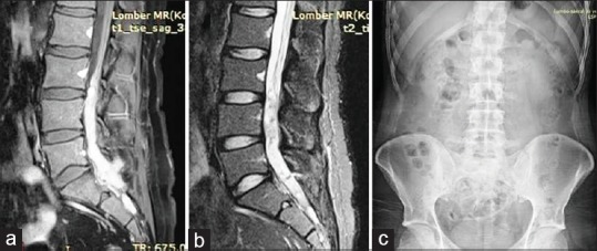
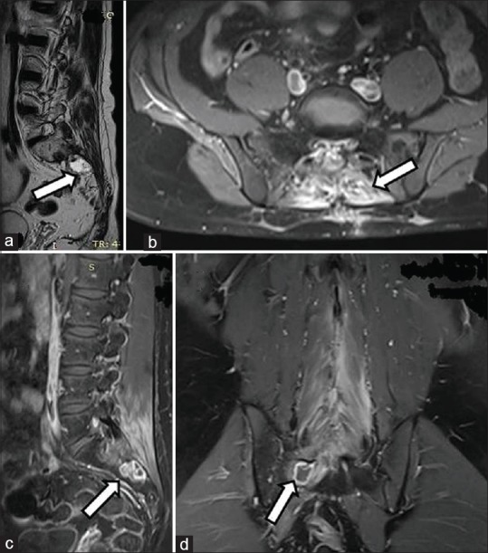
In this study, we evaluated a case of primary spinal oligodendroglioma (PSO) with a rare localization between L3 and S2, and also examined sixty cases in the literature in terms of demographic characteristics, clinical, radiological, and histopathological characteristics, and treatment planning. A case of PSO has been presented, and the relevant literature between 1931 and 2016 was reviewed. A total of 57 papers regarding PSO were found and utilized in this review. The main treatment options include radical surgical excision with neuromonitoring, followed by radiotherapy. Despite these treatment protocols, the relapse rate is high, and treatment does not significantly prolong survival. Oligodendrogliomas are rare among the primary spinal cord tumors. Oligodendrogliomas are predominantly found in the cervical spinal cord, thoracic spinal cord, or junctions during childhood and adulthood. Extension to the sacral region, inferior to the Conus, is very rare. Furthermore, of the sixty cases in the literature, the case we present here is the first to be reported in this particular age group. These localizations usually occur in the pediatric age group and after relapses. While for a limited number of cases the oligodendroglioma initiates in the thoracic region and reaches as far as L2, we encountered a case of an oligodendroglioma within the range of L3 to S2. Clinical findings are observed in accordance with location, and magnetic resonance imaging is the gold standard for diagnosis.

Keywords: Management; primary spinal oligodendroglioma; review.


Conflict of interest statement

There are no conflicts of interest.

Figures

Figure 1
Figure 1
(a) On the T1-weighted magnetic resonance imaging scan with contrast, a dense contrast-enhancing mass is seen spanning from the lower margin of L3 to S2. (b) In the T2-weighted magnetic resonance imaging scan without contrast, a nonhomogenous lesion is seen, including hypointense signal densities. (c) No distinct pathologies were observed on the anterior-posterior X-ray image
Figure 2
Figure 2
(a) After opening the dura, hemorrhagic fragile tumor tissue was seen on the upper tumor margin. (b) As the dura opens distally, fragile tumor tissue can be observed coming away from the intradural space. (c) After the dura is opened and hung from the sides, gray-black-colored tumor tissue is visible between the cauda fibers. (d) An image is shown after the removal of tumor tissues
Figure 3
Figure 3
(a) Basal somatosensory evoked potential (b) basal motor evoked potential (c) dural opening motor evoked potential, and (d) dural opening and motor evoked potential when the tumor area is exposed (e) motor evoked potential during tumor excision is shown (f) closing motor evoked potential is shown (g) exit somatosensory evoked potential and motor evoked potential values are shown
Figure 4
Figure 4
(a and b) The typical circular and flattened appearance for an oligodendroglioma can be observed with uniform round nuclei and sparse or clear cytoplasm with perinuclear halos (a: ×200 and b: ×1000, respectively). (c) Glial fibrillary acidic protein staining showing positive focal immunoreactivity (×200). (d) Nuclear immuno-expression of Ki-67 is shown in a few neoplastic cells (×400)
Figure 5
Figure 5
No pathology was observed in the patient's seeding scans in the (a) T2-weighted sagittal cervical magnetic resonance imaging, (b) T1-weighted sagittal cervical magnetic resonance imaging, (c) T1-weighted sagittal thoracic magnetic resonance imaging; (d) T2-weighted sagittal thoracic magnetic resonance imaging (e) T1-weighted sagittal brain magnetic resonance imaging (f) T1-weighted axial brain magnetic resonance imaging
Figure 6
Figure 6
(a) In the early postoperative period T2-weighted sagittal magnetic resonance imaging scan section without contrast, it can be seen that the tumor tissue has been removed. (b) In the postoperative early period T1-weighted sagittal magnetic resonance imaging scan section without contrast, it can be seen that the tumor tissue has been removed. (c) In the postoperative early period T1-weighted sagittal magnetic resonance imaging scan section with contrast, early postoperative changes can be seen
Figure 7
Figure 7
In the postoperative 6-month (a) T2-weighted sagittal magnetic resonance imaging scan section without contrast, it can be seen that the tumor tissue has been removed (b) T1-weighted sagittal magnetic resonance imaging scan section with contrast, no residue contrast enhancement is observed in the intradural space (c) T1-weighted sagittal magnetic resonance imaging scan section with contrast, contrast enhancement is seen in the sacral region and bone (d) T1-weighted axial magnetic resonance imaging scan section with contrast, contrast enhancement is seen in the sacral region and bone
Figure 8
Figure 8
In the postoperative 15-month (a) T1-weighted sagittal magnetic resonance imaging section without contrast, it can be seen that the tumor tissue sacrum (b) T1-weighted axial magnetic resonance imaging section with contrast, contrast enhancement is observed in the sacrum (c) T1-weighted sagittal magnetic resonance imaging section with contrast, contrast enhancement is seen in the sacral region and bone (d) T1-weighted coronal magnetic resonance imaging section with contrast, contrast enhancement is seen in the sacral region and bone

References

    1. Tunthanathip T, Oearsakul T. Primary spinal cord oligodendroglioma: A case report and review of the literature. Chin Neurosurg J. 2016;2:2.
    1. Yuh WT, Chung CK, Park SH. Primary spinal cord oligodendroglioma with postoperative adjuvant radiotherapy: A case report. Korean J Spine. 2015;12:160–4. - PMC - PubMed
    1. Moorthy NL, Kondeti D, Chander M, Jadhav H, Ashok Spinal cord oligodendroglioma: A case report. IOSR J Dent Med Sci. 2015;14:27–8.
    1. Wang F, Qiao G, Lou X. Spinal cord anaplastic oligodendroglioma with 1p deletion: Report of a relapsing case treated with temozolomide. J Neurooncol. 2011;104:387–94. - PubMed
    1. Guppy KH, Akins PT, Moes GS, Prados MD. Spinal cord oligodendroglioma with 1p and 19q deletions presenting with cerebral oligodendrogliomatosis. J Neurosurg Spine. 2009;10:557–63. - PubMed

Publication types