. 2017 Sep 27:8:1197.
doi: 10.3389/fimmu.2017.01197. eCollection 2017.

Intracellular Crosslinking of Filoviral Nucleoproteins with Xintrabodies Restricts Viral Packaging

Affiliations

Intracellular Crosslinking of Filoviral Nucleoproteins with Xintrabodies Restricts Viral Packaging

Tamarand Lee Darling et al. Front Immunol. .

Abstract

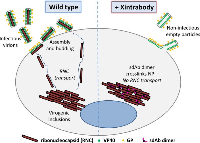
Viruses assemble large macromolecular repeat structures that become part of the infectious particles or virions. Ribonucleocapsids (RNCs) of negative strand RNA viruses are a prime example where repetition of nucleoprotein (NP) along the genome creates a core polymeric helical scaffold that accommodates other nucleocapsid proteins including viral polymerase. The RNCs are transported through the cytosol for packaging into virions through association with viral matrix proteins at cell membranes. We hypothesized that RNC would be ideal targets for crosslinkers engineered to promote aberrant protein-protein interactions, thereby blocking their orderly transport and packaging. Previously, we had generated single-domain antibodies (sdAbs) against Filoviruses that have all targeted highly conserved C-terminal regions of NP known to be repetitively exposed along the length of the RNCs of Marburgvirus (MARV) and Ebolavirus (EBOV). Our crosslinker design consisted of dimeric sdAb expressed intracellularly, which we call Xintrabodies (X- for crosslinking). Electron microscopy of purified NP polymers incubated with purified sdAb constructs showed NP aggregation occurred in a genus-specific manner with dimeric and not monomeric sdAb. A virus-like particle (VLP) assay was used for initial evaluation where we found that dimeric sdAb inhibited NP incorporation into VP40-based VLPs whereas monomeric sdAb did not. Inhibition of NP packaging was genus specific. Confocal microscopy revealed dimeric sdAb was diffuse when expressed alone but focused on pools of NP when the two were coexpressed, while monomeric sdAb showed ambivalent partition. Infection of stable Vero cell lines expressing dimeric sdAb specific for either MARV or EBOV NP resulted in smaller plaques and reduced progeny of cognate virus relative to wild-type Vero cells. Though the impact was marginal at later time-points, the collective data suggest that viral replication can be reduced by crosslinking intracellular NP using relatively small amounts of dimeric sdAb to restrict NP packaging. The stoichiometry and ease of application of the approach would likely benefit from transitioning away from intracellular expression of crosslinking sdAb to exogenous delivery of antibody. By retuning sdAb specificity, the approach of crosslinking highly conserved regions of assembly critical proteins may well be applicable to inhibiting replication processes of a broad spectrum of viruses.

Keywords: Ebola; Marburg; VHH; crosslinker; intrabody; nucleoprotein; single-domain antibodies; virus-like particle.


Figures

Figure 1
Figure 1
Overall hypothesis of the approach showing that crosslinking a viral structural protein within the cell using a multimeric single-domain antibody (sdAb) will impede viral replication by disrupting the orderly assembly of infectious virus particles (virions). The dimeric sdAb can be introduced into the uninfected target cell or virus-infected cell via endogenous gene expression, gene delivery, protein transfection, or protein transduction to mediate the antiviral activity. Here, we focus on utilizing Marburgvirus and Ebolavirus nucleoprotein (NP) as our model target since we have sdAb to hand that bind polyvalent assemblies. Our working theory is that our sdAb dimers or Xintrabodies are crosslinking NP epitopes among ribonucleocapsids (RNCs) within viral factories and inhibiting their transport to the cell membrane for further assembly into virions. It should be noted that other antigens involved in assembly may be equally effective provided suitable sdAb are available, although targeting antigens that occur in viral factories may be advantageous owing to high local concentrations. The overall approach may be applicable to many other viruses and scaffolding components and leverages relatively small amounts of minute 30 kDa dimeric sdAb crosslinking MDa sized targets to impede productive viral replication.
Figure 2
Figure 2
Purification of single-domain antibody (sdAb) proteins and recombinant nucleoprotein (NP) for ELISA characterization. Coomassie stained SDS-PAGE gel showing 5 µg of each sdAb monomer (A) and dimer (B) purified from Escherichia coli periplasm. (C) Silver stained SDS-PAGE gel of Marburgvirus (MARV) (M) and Ebolavirus (EBOV) (Z) NP preparations following large-scale transient transfection and purification through centrifugation steps and banding on CsCl gradients. (D) ELISA titration of the anti-MARV sdAb monomers and dimers over MARV NP with the highest concentration also applied to Bundibugyo NP (this was expressed at higher levels than Zaire NP and so was convenient to use for controls yet shares high homology at the C-terminal domain for our studies). (E) ELISA titration of anti-EBOV sdAb E monomer and dimer over EBOV NP and MARV NP.
Figure 3
Figure 3
Evaluating expression of various single-domain antibody (sdAb) formats as intrabodies within HEK 293T cells. (A) Mammalian expression vector puma1 built for these studies utilizes a human cytomegalovirus immediate early gene enhancer and promoter (CMV IE) and the adenovirus tripartite 5′-non-coding region with a hybrid splice donor acceptor with other components from the pcDNA stable (Invitrogen) including high copy number in both Escherichia coli and HEK 293T cells and G418 resistance cassette for stable line generation. (B) Anti-C9 Western blot of whole cell lysates of HEK 293T cells transiently transfected with C9 tagged wild-type llama sdAb sequences or human optimized sequences identify those clones with low and high relative expression levels. Cells were harvested 72 h post-transfection. (C) Anti-C9 Western blot of HEK 293T soluble (including cytosol) and insoluble (including membranes) fractions following transient expression of llama sdAb as monomeric and dimeric versions. (D) Coomassie blue stained SDS-PAGE of the soluble fractions reveal visible bands at the expected places for sdAb B and E monomers and just visible are the corresponding dimers.
Figure 4
Figure 4
Examining the ability of single-domain antibody (sdAb) monomers and dimers to crosslink nucleoprotein (NP) in vitro by electron microscopy. 10:1 M ratios of the anti-Marburgvirus (MARV) monomer (M1) or dimer (M2), or anti-Ebolavirus (EBOV) monomer (E1) or dimer (E2) were combined with MARV or EBOV NP and equilibrated for 1 h prior to transmission microscopy. In the absence of crosslinking the individual helical filaments of NP, polymers are visible in the 100,000× images for both MARV and EBOV NP.
Figure 5
Figure 5
Quantification of the 100,000× images from Figure 4 using Cell Profiler to reveal the extent of aggregated and non-aggregated nucleoprotein (NP).
Figure 6
Figure 6
Exploring the impact of coexpressing the anti-Marburgvirus (MARV) single-domain antibody (sdAb) monomer (M1) or dimer (M2), or anti-Ebolavirus (EBOV) monomer (E1) or dimer (E2) on nucleoprotein (NP) incorporation into VLP. (A) Sliver stained SDS-PAGE analysis of crude VLP preparations following coexpression of MARV NP and/or VP40 with the various sdAb monomers and dimers. (B) Coomassie stained SDS-PAGE analysis of cell lysates stemming from the VLP production in panel (A). (C) Sliver stained SDS-PAGE analysis of crude VLP preparations following coexpression of EBOV NP and/or VP40 with the various sdAb monomers and dimers. (D) Coomassie stained SDS-PAGE analysis of cell lysates stemming from the VLP production in panel (C).
Figure 7
Figure 7
Localization of single-domain antibody (sdAb) monomer or dimer when coexpressed with nucleoprotein (NP) and VP40 and the impact on NP-VP40 colocalization. Immunofluorescence microscopy of transiently cotransfected Vero E6 cells producing anti-Marburgvirus (MARV) sdAb monomer (M1) or dimer (M2), or anti-Ebolavirus (EBOV) sdAb monomer (E1) or dimer (E2) with either (A) MARV VP40 and MARV NP genes or (B) EBOV VP40 and NP genes.
Figure 8
Figure 8
Production and distribution of single-domain antibody (sdAb) dimers within the Vero E6 based stable cell lines. (A) Western blot of stable cell lines expressing the anti-Marburgvirus (MARV) dimer (Vero-M2) or anti-Ebolavirus (EBOV) dimer (Vero-E2) and probing for antibody via the C9 tag or for β-actin. (B) Staining of parent (Vero-wt) and stable cell lines Vero-M2 and Vero-E2 for the distribution of antibody via the C9 tag. (C) Transient expression of MARV HA-NP within the parental and transgenic cell lines demonstrates the ability of the anti-MARV dimer but not the anti-EBOV dimer to colocalize with nucleoprotein (NP) puncta. (D) Transient expression of EBOV HA-NP within the parental and transgenic cell lines demonstrates the ability of the anti-EBOV dimer but not the anti-MARV dimer to colocalize with NP puncta.
Figure 9
Figure 9
Impact of Xintrabodies on viral replication. Marburgvirus (MARV) (A) and Ebolavirus (EBOV) (B) were titrated on wild-type Vero cells (wt) or the stable lines expressing the anti-MARV dimer (M2) or anti-EBOV dimer (E2) and plaque formation under semi-solid overlay allowed to proceed for 10 days. The experiment was performed twice for MARV and three times for EBOV and error bars represent the SD. Diameters of approximately 150 plaques from the titrations were measured using ImageJ measure analysis tool and plotted for MARV (C) and EBOV (D). An unpaired, one-tail t-test analysis revealed a p-value of <0.0001 (****) while ns indicates no significant difference. Bars indicate SD and the mean. (E) MARV and EBOV were used to infect wild-type or cognate single-domain antibody dimer stable cell lines in a growth kinetics format with liquid supernatant collections at the times indicated. Plaque titrations of the supernatants were performed on wild-type cells and results are graphed as a percentage of titers resulting from the wild-type time-course. The experiment was performed three times for MARV and twice for EBOV. An unpaired, one-tail t-test was conducted using Excel, * indicating a p-value <0.05 and ** indicating a p-value <0.005.

References

    1. Marasco WA. Intrabodies: turning the humoral immune system outside in for intracellular immunization. Gene Ther (1997) 4:11–5.10.1038/sj.gt.3300346 - DOI - PubMed
    1. Baltimore D. Gene therapy. Intracellular immunization. Nature (1988) 335:395–6.10.1038/335395a0 - DOI - PubMed
    1. Antman KH, Livingston DM. Intracellular neutralization of SV40 tumor antigens following microinjection of specific antibody. Cell (1980) 19:627–35.10.1016/S0092-8674(80)80039-0 - DOI - PubMed
    1. Fujimoto Y, Ozaki K, Maeda M, Nishijima K, Takakuwa H, Otsuki K, et al. Resistance to influenza A virus infection in transformed cell lines expressing an anti-PB2 monoclonal antibody. Vet J (2013) 198:487–93.10.1016/j.tvjl.2013.09.019 - DOI - PubMed
    1. Kontermann RE. Intrabodies as therapeutic agents. Methods (2004) 34:163–70.10.1016/j.ymeth.2004.04.002 - DOI - PubMed

LinkOut - more resources