. 2017 Sep 27;3(9):e00416.
doi: 10.1016/j.heliyon.2017.e00416. eCollection 2017 Sep.

Placental extract ameliorates non-alcoholic steatohepatitis (NASH) by exerting protective effects on endothelial cells

Affiliations

Placental extract ameliorates non-alcoholic steatohepatitis (NASH) by exerting protective effects on endothelial cells

Akihiro Yamauchi et al. Heliyon. .

Abstract

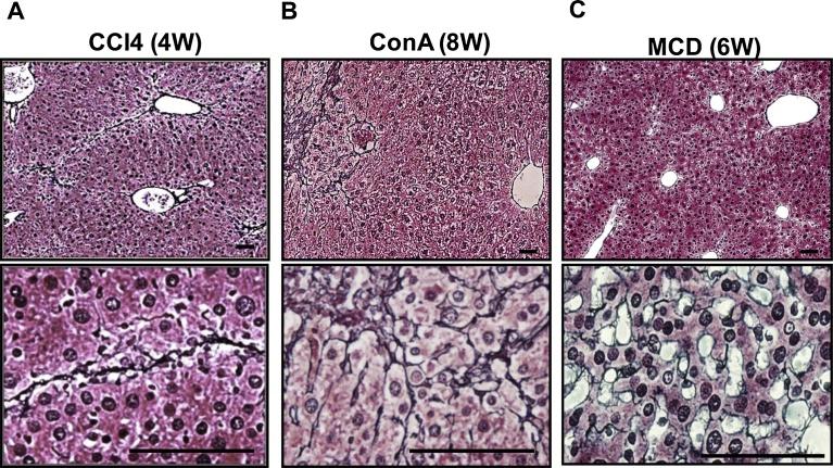
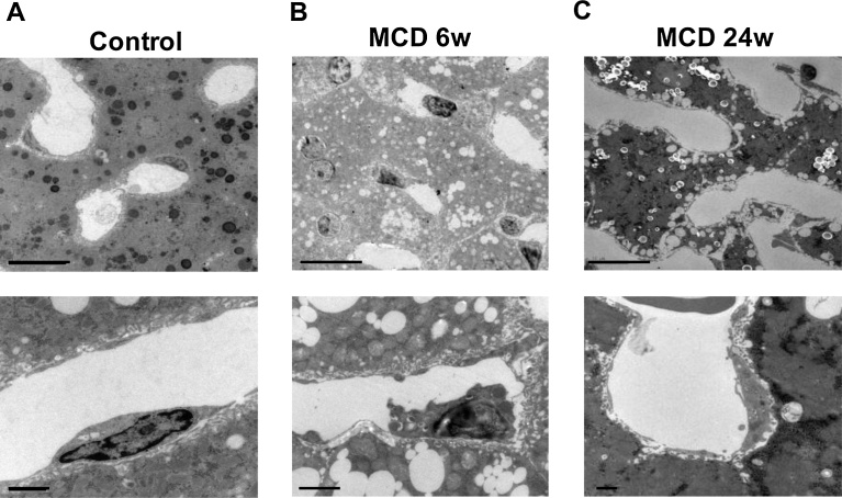
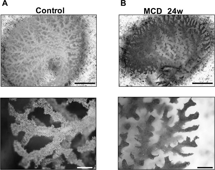
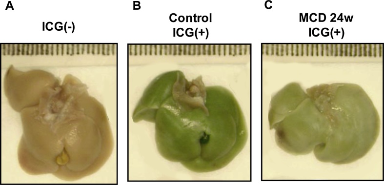
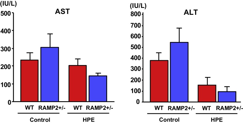
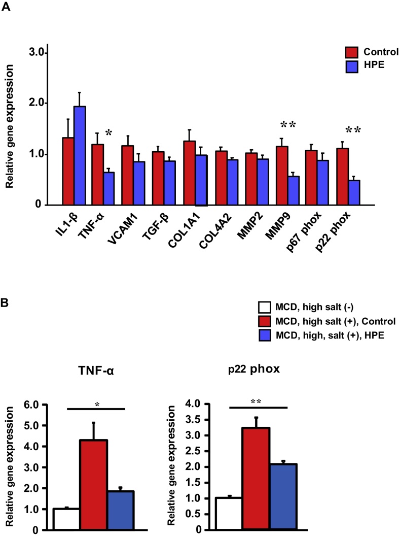
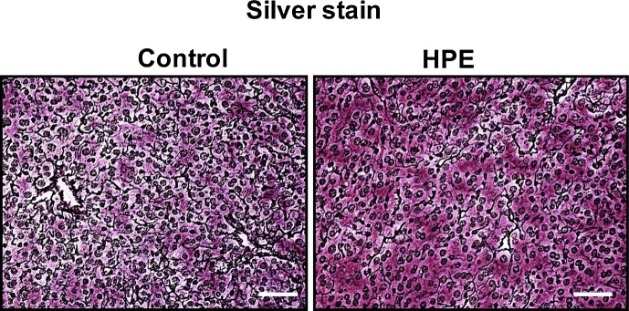
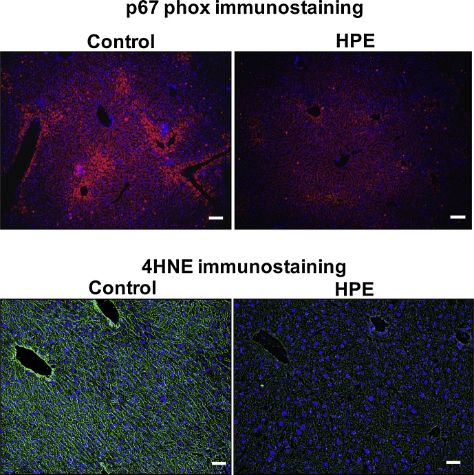
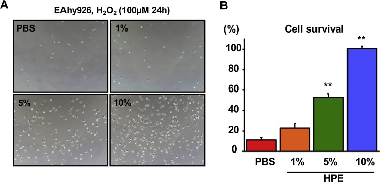
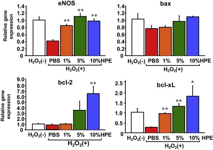
Non-alcoholic steatohepatitis (NASH) is a severe form of fatty liver disease that is defined by the presence of inflammation and fibrosis, ultimately leading to cirrhosis and hepatocellular carcinoma. Treatment with human placental extract (HPE) reportedly ameliorates the hepatic injury. We evaluated the effect of HPE treatment in a mouse model of NASH. In the methione- and choline-deficient (MCD) diet-induced liver injury model, fibrosis started from regions adjacent to the sinusoids. We administered the MCD diet with high-salt loading (8% NaCl in the drinking water) to mice deficient in the vasoprotective molecule RAMP2 for 5 weeks, with or without HPE. In both the HPE and control groups, fibrosis was seen in regions adjacent to the sinusoids, but the fibrosis was less pronounced in the HPE-treated mice. Levels of TNF-α and MMP9 expression were also significantly reduced in HPE-treated mice, and oxidative stress was suppressed in the perivascular region. In addition, HPE dose-dependently increased survival of cultured endothelial cells exposed to 100 μM H2O2, and it upregulated expression of eNOS and the anti-apoptotic factors bcl-2 and bcl-xL. From these observations, we conclude that HPE ameliorates NASH-associated pathologies by suppressing inflammation, oxidative stress and fibrosis. These beneficially effects of HPE are in part attributable to its protective effects on liver sinusoidal endothelial cells. HPE could thus be an attractive therapeutic candidate with which to suppress progression from simple fatty liver to NASH.

Keywords: Health Sciences; Metabolism; Pathology; Pharmaceutical science.


Figures

Fig. 1
Fig. 1
MCD diet caused fibrosis adjacent to the liver sinusoids. C57BL/6J wild-type mice were used for chronic liver injury models. Shown are silver-stained liver sections at lower (upper panels) and higher (lower panels) magnification. Silver-stained fibrotic areas were detected mainly between the parenchymal hepatocytes in CCl4-treated (A) and Con A-treated (B) mice, but were detected adjacent to the sinusoids in mice fed the MCD diet (C). Scale bars = 100 μm.
Fig. 2
Fig. 2
The MCD diet caused damage to LSECs. Electron microscopic observation of the liver sinusoids from control mice (A) and mice fed the MCD diet for 6 weeks (B) or 24 weeks (C). Upper and lower panels show lower and higher magnifications, respectively. The MCD diet caused fatty liver changes, characterized by the accumulation of lipid droplets within the hepatocytes (upper panels in B and C) as well as cellular deformities (lower panel in B) and nuclear loss (lower panel in C) in LSECs. Scale bars = 10 μm in upper panels, 1 μm in lower panels.
Fig. 3
Fig. 3
Vascular casting showed the MCD diet collapsed the structure of sinusoids. Acrylic resin casting of the liver vasculature from a control mouse (A) and a mouse fed the MCD diet for 24 weeks (B). Upper and lower panels show lower and higher magnifications, respectively. In control mice, sinusoidal casts were well visualized to the tip of each vascular branch (lower panel in A). In MCD-treated mice, the casting of the smaller vessels was indiscernible (lower panel in B). Scale bars = 5 mm in upper panels, 1 mm in lower panels.
Fig. 4
Fig. 4
Indocyanine green (ICG) uptake was diminished in the livers of mice fed the MCD diet. Shown is the appearance of the liver without ICG injection (A) and after intravenous injection of ICG in control and (B) MCD-treated mice (C). Uptake of ICG was diminished in the MCD-treated mice (compare B and C).
Fig. 5
Fig. 5
HPE treatment ameliorated liver injury. Serum AST and ALT levels in heterozygous RAMP2 knockout mice (RAMP2+/−) and wild-type mice administered the MCD diet for 5 weeks with 8% NaCl in the drinking water. Mice were treated with intramuscular injections of saline (Control) or HPE. Bars are means ± SEM. n = 8 in each group. Serum transaminases tended to be higher in the Control groups than in the HPE-treated groups.
Fig. 6
Fig. 6
HPE treatment suppressed expression of inflammation-, fibrosis- and oxidative stress-related genes. A. Relative expression of the indicated genes in the livers of control and HPE-treated RAMP2+/− mice. The median of the control group was assigned a value of 1. Bars are means ± SEM. n = 8 in each group. *p < 0.05, **p < 0.01. HPE suppressed expression of inflammation-, fibrosis- and oxidative stress-related genes. B. Relative gene expression of TNF-α and p22phox in the livers of RAMP2+/− mice. The mean of the MCD, high salt (−) group was assigned a value of 1. Bars are means ± SEM. n = 8 in each group. *p < 0.05, **p < 0.01. Compared to the MCD, high salt (−) group, the MCD, high salt (+) group showed significantly elevated expression of both TNF-α and p22phox. HPE-treatment in MCD, high salt (+) group partially normalized expression of the inflammatory and oxidative stress markers.
Fig. 7
Fig. 7
HPE treatment suppressed fibrotic changes in the livers of mice fed the MCD diet. Shown are sliver stained liver sections from representative control and HPE-treated mice. Silver stained fibrotic areas were detected adjacent to the sinusoids in both groups, but the areas were smaller in HPE-treated mice. Scale bars = 100 μm.
Fig. 8
Fig. 8
HPE-treatment suppressed oxidative stress in the livers of mice fed the MCD diet. Shown is immunostaining for p67 phox, a NADPH oxidase subunit (upper panels) and 4HNE, a lipid peroxidation product (lower panels), in liver sections. Immunostaining of p67 phox and 4HNE was detected in the perivascular regions of both control and HPE-treated mice, but it was much weaker in the HPE-treated group. Scale bars = 100 μm.
Fig. 9
Fig. 9
HPE treatment increased survival among cultured endothelial cells. EAhy926 human endothelial cells were exposed to 100 μM H2O2 for 24 h with or without HPE-treatment. Photos of the cultured endothelial cells (A) and the comparison of the cell survival (B) are shown. Bars are means ± SEM. The 10% HPE-treated group was assigned a value of 100%. n = 8 in each group. **p < 0.01. HPE dose-dependently increased endothelial cell survival.
Fig. 10
Fig. 10
HPE-treatment upregulated expression of eNOS and anti-apoptotic genes. Expression of the indicated genes in cultured EAhy926 human endothelial cells. Bars are means ± SEM. The mean of the control H2O2(−) group was assigned a value of 1. n = 8 in each group. *p < 0.05, **p < 0.01. HPE-treatment upregulated expression of eNOS, reflecting increased endothelial cell viability. HPE-treatment also upregulated expression of the anti-apoptotic factors bcl-2 and bcl-xL, while expression of the pro-apoptotic factor bax was unchanged.

References

    1. Arab J.P., Candia R., Zapata R., Munoz C., Arancibia J.P., Poniachik J. Management of nonalcoholic fatty liver disease: an evidence-based clinical practice review. World J. Gastroenterol. 2014;20:12182–12201. - PMC - PubMed
    1. Arai T., Sakurai T., Kamiyoshi A., Ichikawa-Shindo Y., Iinuma N., Iesato Y. Induction of LYVE-1/stabilin-2-positive liver sinusoidal endothelial-like cells from embryoid bodies by modulation of adrenomedullin-RAMP2 signaling. Peptides. 2011;32:1855–1865. - PubMed
    1. Caldwell-Kenkel J.C., Currin R.T., Tanaka Y., Thurman R.G., Lemasters J.J. Reperfusion injury to endothelial cells following cold ischemic storage of rat livers. Hepatology. 1989;10:292–299. - PubMed
    1. Chen C.P., Tsai P.S., Huang C.J. Antiinflammation effect of human placental multipotent mesenchymal stromal cells is mediated by prostaglandin E2 via a myeloid differentiation primary response gene 88-dependent pathway. Anesthesiology. 2012;117:568–579. - PubMed
    1. Day C.P., James O.F. Steatohepatitis: a tale of two hits. Gastroenterology. 1998;114:842–845. - PubMed