Review
. 2018 Jun;9(3):215-222.
doi: 10.1007/s12975-017-0572-0. Epub 2017 Oct 11.

Dysfunction of Membrane Trafficking Leads to Ischemia-Reperfusion Injury After Transient Cerebral Ischemia

Affiliations
Review

Dysfunction of Membrane Trafficking Leads to Ischemia-Reperfusion Injury After Transient Cerebral Ischemia

Dong Yuan et al. Transl Stroke Res. 2018 Jun.

Abstract

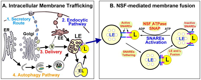
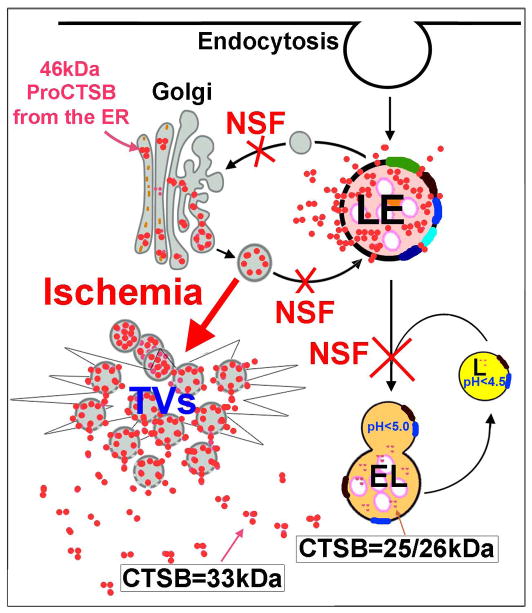
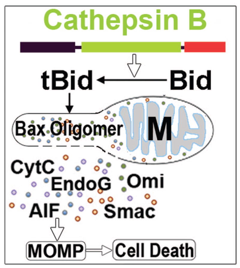
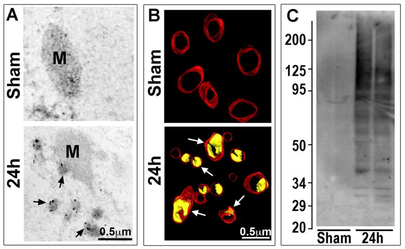
Neurons require an extraordinarily high level of membrane trafficking activities because of enriched axonal terminals and dendritic branches. For that reason, defects in the membrane trafficking pathway are a hallmark of most, and may be all, neurodegenerative disorders. A major cellular membrane trafficking pathway is the Golgi apparatus (Golgi hereafter)-late endosome-lysosome axis for supplying lysosomal enzymes. This pathway is regulated by N-ethylmaleimide-sensitive factor (NSF) ATPase. This review article is to discuss a novel hypothesis that brain ischemia inactivates NSF ATPase, resulting in a cascade of events of disruption of the Golgi-endosome-lysosome pathway, release of cathepsin B (CTSB), and induction of mitochondrial outer membrane permeabilization (MOMP) during the postischemic phase. This hypothesis is supported by recent studies demonstrating that NSF is trapped into inactive protein aggregates in neurons destined to die after brain ischemia. Consequently, Golgi, transport vesicles (TVs), and late endosomes (LEs) are accumulated and damaged, which is followed by CTSB release from these damaged structures. Moderate release of CTSB cleaves Bax-like BH3 protein (Bid) to become active truncated Bid (tBid). Active tBid is then translocated to the mitochondrial outer membrane, resulting in oligomerization of BCL2-associated X protein (Bax) forming the mitochondrial outer membrane pores, and releasing mitochondrial intramembranous proteins. Extensive CTSB release, however, can digest cellular proteins indiscriminately to induce cell death. Based on these new observations, we propose a novel hypothesis, i.e., brain ischemia leads to NSF inactivation, resulting in a massive buildup of damaged Golgi, TVs and LEs, fatal release of CTSB, induction of MOMP, and eventually brain ischemia-reperfusion injury.

Keywords: Brain ischemia-reperfusion injury; Golgi; Late endosome; Lysosome; Membrane trafficking; Mitochondrial outer membrane permeabilization; NSF, cathepsin B; Transport vesicle.


Figures

Fig. 1
Fig. 1
A: Cellular membrane trafficking encompasses 4 major types: (1) secretory or exocytic route, (2) endocytic pathway; (3) newly synthesized biomolecules delivery; and (4) autophagy pathway (Fig. 1A). ER = endoplasmic reticulum. V = transport vesicle. LE = late endosome. EE = early endosome. EL = endolysosome. AP = autophagosome. L = lysosome. B: Example of LE and lysosomal membrane fusion in the inset of A. Membrane fusion is mediated by active SNAREs tethering to bring two membranes together. After membrane fusion, SNAREs form inactive complexes and must be regenerated by NSF ATPase for the next round of fusion. During this process, NSF ATPase binds to inactive SNAREs via a SNAP adaptor to hydrolyze adenosine triphosphate (ATP) to adenosine diphosphate (ADP) and inorganic phosphate for providing the energy. Brain ischemia leads to deposition NSF into inactive protein aggregates, thus stopping all membrane trafficking activities in postischemic neurons.
Fig. 2
Fig. 2
Confocal microscopic images of CA1 neurons labeled with NSF antibody. Brain sections were obtained from a sham-operated control rat and rat subjected to 15 min of ischemia followed by 24 h of reperfusion. NSF is located in the peri-nuclear and dendritic truck (Sham, arrowheads), as well as in the neuropil (Sham, arrows) of sham-operated control CA1 neurons. NSF is mostly depleted from the CA1 neuronal perinuclear region and dendritic trunk (24h, arrowheads), but remains relatively at the sham control level in the CA1 neuropil (24h, arrows) at 24 h of reperfusion after transient cerebral ischemia.
Fig. 3
Fig. 3
EM micrographs of hippocampal CA1 neurons stained with uranium-lead. CA1 tissue sections were obtained from rats subjected to non-ischemic sham surgery or 20 min of ischemia followed by 24 h of reperfusion. a: A sham neuron shows normal rough endoplasmic reticulum (ER), ribosomal rosettes (small arrows), mitochondria (M), Golgi apparatus (G), late endosome (large arrow), and endolysosome (EL, also known as secondary lysosome). b: A 24 h reperfused neuron shows accumulation of Golgi fragments (Gf), transport vesicles (arrowheads), enlarged late endosome (large arrows), monoribosomes (small arrows), and ribosomal aggregates (stars). c and d: Higher magnifications of the insets of “a and b” show that ELs and a LE (arrow) from the sham control neuron has intact lipid membranes, whereas an enlarged LE from the postischemic neuron has a number of membrane break damage (arrowheads). Scale bar = 0.5 μm.
Fig. 4
Fig. 4
CTSB has three active forms: (i) 46 kDa Golgi CTSB located in Golgi/TVs, (ii) 33 kDa CTSB in late endosome (LE), and (iii) 24/25 kDa lysosome CTSB in endolysosome (EL) and lysosome (L).
Fig. 5
Fig. 5
Inactivation of NSF ATPase after brain ischemia leads to a cascade of events of massive buildup of Golgi fragments, TVs and enlarged LEs, fatal release of 33 kDa CTSB, and brain ischemia-reperfusion injury.
Fig. 6
Fig. 6
CTSB release induces mitochondrial outer membrane permeabilization (MOMP): Released CTSB converts Bid to active truncated Bid (tBid), and leads tBid and Bax mitochondrial (M) translocation and Bax oligomerization. Bax oligomerization forms mitochondrial outer membrane pore, resulting in MOMP. Consequently, mitochondrial intermembrane space proteins are released into the cytoplasm, eventually resulting in neuronal death after brain ischemia. CytC = cytochrome C. AIF = apoptosis inducing factor. EndoG = endonuclease G. HtrA2/Omi = a proapoptotic mitochondrial serine protease. SMAC/Diablo = Second mitochondria-derived activator of caspase/direct inhibitor of apoptosis-binding protein with low pI.
Fig. 7
Fig. 7
A: Ubiquitin immunogold EM micrographs of mitochondrion (M). EM micrographs of CA1 neurons from a sham-operated non-ischemic control rat and a rat subjected to 15 min ischemia followed by 24h of reperfusion. Ubiquitin immunogold labels mitochondrion and ubiquitinated protein aggregates at 24h of reperfusion (arrows). B: EM three-dimensional reconstruction of CA1 neuronal mitochondria in brain sections from sham-operated control and 24h reperfused rats. The red indicates the mitochondrial outer membrane. The yellow (arrows) represents the vacuole structures formed between the inner- and outer-mitochondrial membranes after brain ischemia. Scale bars in A and B = 0.5 μm. C: Western blotting of ubiquitin in the samples of detergent-salt insoluble mitochondrial pellets from sham-operated rats and rats subjected to 15 min of ischemia followed by 24 h of reperfusion. The molecular size is shown on the left.

References

    1. Smith ML, Auer RN, Siesjo BK. The density and distribution of ischemic brain injury in the rat following 2–10 min of forebrain ischemia. Acta Neuropathol (Berl) 1984;64:319–332. - PubMed
    1. Taniguchi D, Baernstein A, Nichol G. Cardiac arrest: a public health perspective. Emerg Med Clin North Am. 2012 Feb;30(1):1–12. - PubMed
    1. Kirino T, Tamura A, Sano K. Delayed neuronal death in the rat hippocampus following transient forebrain ischemia. Acta Neuropathol. 1984;64:139–147. - PubMed
    1. Kirino T, Sano K. Fine structural nature of delayed neuronal death following ischemia in the gerbil hippocampus. Acta Neuropathol. 1984;62:209–218. - PubMed
    1. Lin B, Ginsberg MD, Busto R. Hyperglycemic exacerbation of neuronal damage following forebrain ischemia: microglial, astrocytic and endothelial alterations. Acta Neuropathol. 1998;96:610–620. - PubMed

Publication types

MeSH terms

Substances