Review
. 2017 Oct 12:3:17073.
doi: 10.1038/nrdp.2017.73.

Syphilis

Affiliations
Review

Syphilis

Rosanna W Peeling et al. Nat Rev Dis Primers. .

Abstract

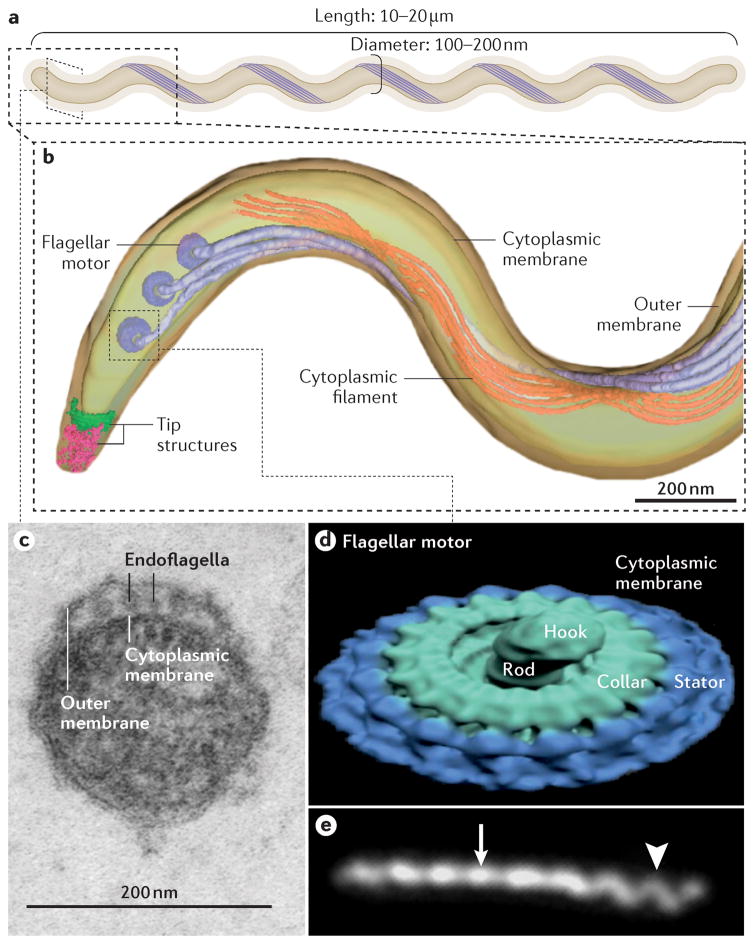
Treponema pallidum subspecies pallidum (T. pallidum) causes syphilis via sexual exposure or via vertical transmission during pregnancy. T. pallidum is renowned for its invasiveness and immune-evasiveness; its clinical manifestations result from local inflammatory responses to replicating spirochaetes and often imitate those of other diseases. The spirochaete has a long latent period during which individuals have no signs or symptoms but can remain infectious. Despite the availability of simple diagnostic tests and the effectiveness of treatment with a single dose of long-acting penicillin, syphilis is re-emerging as a global public health problem, particularly among men who have sex with men (MSM) in high-income and middle-income countries. Syphilis also causes several hundred thousand stillbirths and neonatal deaths every year in developing nations. Although several low-income countries have achieved WHO targets for the elimination of congenital syphilis, an alarming increase in the prevalence of syphilis in HIV-infected MSM serves as a strong reminder of the tenacity of T. pallidum as a pathogen. Strong advocacy and community involvement are needed to ensure that syphilis is given a high priority on the global health agenda. More investment is needed in research on the interaction between HIV and syphilis in MSM as well as into improved diagnostics, a better test of cure, intensified public health measures and, ultimately, a vaccine.


Conflict of interest statement

Competing interests

J.D.R. receives royalties for licensing of syphilis diagnostics reagents. The other authors declare no competing interests.

Figures

Figure 1
Figure 1. Treponema pallidum
A | Like all spirochetes, T. pallidum consists of a protoplasmic cylinder and cytoplasmic membrane bounded by a thin peptidoglycan sacculus and outer membrane,. Usually described as spiral-shaped, T. pallidum is actually a thin planar wave similar to Borrelia burgdorferi, the agent of Lyme borreliosis. The bacterium replicates slowly and poorly tolerates desiccation, elevated temperatures and high oxygen tensions. B | Periplasmic flagellar filaments, a defining morphological feature of spirochetes, originate from nanomotors situated at each pole and wind around the cylinder atop the peptidoglycan, overlapping at mid-cell. Force exerted by the rigid filaments against the elastic peptidoglycan deforms the sacculus to create the flat wave morphology of the spirochete. Panel B used with permission from Ref. C | Ultra-thin section of T. pallidum showing the outer and cytoplasmic membranes and flagellar filaments (endoflagella) within the periplasmic space. D | Surface rendering of a flagellar motor based on cryoelectron tomograms. Panel D used permission from Ref.. E | Darkfield micrograph showing the flat-wave morphology of Tpallidum. The arrow and arrowhead indicate segments that are oriented 90° from each other. The different appearances of the helical wave at 90° to the viewer can be explained only by a flat wave morphology; a corkscrew would appear the same from any angle. Panel E used permission from Ref .
Figure 2
Figure 2. Incidence of syphilis worldwide
The WHO estimates of incident cases of syphilis by region in 2012 are shown for the different geographical regions. Data from Ref.
Figure 3
Figure 3. Molecular architecture of the cell envelope of Treponema pallidum
Shown in the outer membrane are TP0751 (as known as pallilysin), and Tpp17 (also known as TP0435), — two surface-exposed lipoproteins; TP0453, a lipoprotein attached to the inner leaflet of the outer membrane; BamA (also known as TP0326),; a full-length T. pallidum repeat (Tpr) attached by its N-terminal portion to the peptidoglycan,; and a generic β-barrel that represents other non-Tpr outer-membrane proteins identified by computational mining of the T. pallidum genome. Substrates and nutrients present in high concentration in the extracellular milieu (such as, glucose) traverse the outer membrane through porins, such as TprC. At the cytoplasmic membrane, prototypic ABC-like transporters (such as RfuABCD, a riboflavin transporter) use a periplasmic substrate-binding protein (SBP), usually lipoproteins, and components with transmembrane and ATP-binding domains to bind nutrients that have traversed the outer membrane for transport across the cytoplasmic membrane. The energy coupling factor (ECF)-type ABC transporters use a transmembrane ligand-binding protein in place of a separate periplasmic SBP for binding of ligands (BioMNY is thought to transport biotin). Symporter permeases (for example, TP0265) use the chemiosmotic or electrochemical gradient across the cytoplasmic membrane to drive substrate transport. The tripartite ATP-independent periplasmic (TRAP)-type transporters also use transmembrane electrochemical gradients to drive substrate transport; the periplasmic component protein TatT (also known as TP0956) likely associates with the SBP TatP (also known as TP0957) that binds ligands (perhaps hydrophobic molecules, such as long chain fatty acids), uptake of which is probably is facilitated by the permease TatQ-M (also known as TP0958) ,. Figure adapted from Ref. with permission.
Figure 4
Figure 4. Treponema pallidum invasion
A | Transmission electron micrograph of T. pallidum (arrowheads) penetrating the junctions between cultured umbilical vein endothelial cells. ‘Inter-junctional invasion’ following attachment to vascular endothelium is thought to provide T. pallidum access to tissue parenchyma during haematogenous dissemination. Reprinted with permission from Reference. B | Immunohistochemical staining (using commercial anti-T. pallidum antibodies) of a secondary syphilis skin lesion reveals abundant spirochetes embedded within a mixed cellular inflammatory infiltrate in the papillary dermis. The inflammatory response elicited by spirochetes replicating in tissues is widely thought to be the cause of clinical manifestations in all stages of syphilis. Reprinted with permission from. C | Human syphilitic serum (HSS) dramatically enhances opsonophagocytosis of T. pallidum by purified human peripheral blood monocytes compared with D | normal human serum (NHS). Arrowheads indicate treponemes being degraded within phagolysosomes.
Figure 5
Figure 5. Clinical presentation of primary, secondary and congenital syphilis
A | Primary chancre. B | Primary chancre with rash of secondary syphilis. C | Secondary syphilis in a pregnant woman, who has palmar rash. D | Secondary syphilis as palmar rash. E | 3-month old baby with congenital syphilis, showing hepatosplenomegaly and desquamating rash. The child also presented with nasal discharge. F | Typical palmar desquamating rash in baby with congenital syphilis.
Figure 6
Figure 6. Serological response to primary and secondary syphilis
Diagnosis of syphilis can be made by measuring a patient’s serological response to infection. IgM antibodies against Treponema pallidum proteins are the first to appear, followed a few weeks later by IgG antibodies. Both IgM and IgG antibodies can be measured using treponemal tests such as the T. pallidum Haemagglutination Assay (TPHA), T. pallidum Particle Assay (TPPA), Fluorescent Treponemal Antibody Absorption assay (FTA-ABS), enzyme immunoassays (EIA) and Chemilluminescent immunoassays (CIA). IgM and IgG antibodies against proteins that are not specific to T. pallidum (non-treponemal antibodies) can be detected using the Rapid Plasma Reagin (RPR), Venereal Disease Research Laboratory (VDRL) or toluidine red unheated serum (TRUST) tests and usually appear 2–3 week after treponemal antibodies. With effective treatment (which is arbitrarily shown here at 6 months), the non-treponemal antibody levels decline whereas the treponemal antibodies remain high for many years. In ~20% of patients, non-trepnemal antibodies persist 6 months after treatment; these individuals are labelled as having a serofast status. Despite repeated treatment, ~11% of patients remain serofast. Here, we show early syphilis (including primary, secondary and early latent infections; infectious syphilis) and late syphilis (including late latent and tertiary infections) as being ≤1 year in duration and >1 year in duration, respectively, in line with US and European guidelines. However, the WHO guidelines place this demarcation at 2 years. Beyond primary and secondary syphilis, the pattern of serological response over time is less well defined and is accordingly not shown.
Figure 7
Figure 7. Screening algorithms for syphilis
A | The traditional algorithm begins with a qualitative non-treponemal test (NTT) that is confirmed with a treponemal test (TT). This algorithm has a high positive predictive value when both tests are reactive, although early primary and previously treated infections can be missed owing to the lower sensitivity of NTTs. Importantly, this algorithm is less costly than reverse screening algorithms, and does not require highly specialized laboratory equipment, but is limited by subjective interpretation of the technologist. Additionally, false negative NTT results can arise from the prozone effect (when there is an excess of antibody). Finally, because the traditional algorithm is not always followed by a confirmatory TT, previously treated, early untreated and late latent cases can be missed and biologically false-positive cases can be overtreated. B | The reverse screening algorithm uses a TT with recombinant T. pallidum antigens in enzyme immunoassay (EIA) or chemiluminescence immunoassay (CIA) formats that, when reactive, is followed by an NTT. This approach is associated with higher initial setup costs and ongoing operational costs than the traditional algorithm, but the algorithm permits treatment of 99% of syphilis cases, compared to the traditional algorithm in a low-prevalence setting. Also, because TTs are not flocculation assays, false negative tests due to the Prozone effect do not occur. However, in high-risk populations, screening with a TT can result in a high rate of positive results due to previously treated infections, leading to increased clinician workload needed to review cases and determine appropriate management. Some guidelines recommend further evaluation of reactive TT with a quantitative NTT and, if results of the latter are nonreactive, a second (different) TT to help resolve the discordant results,,. The European Centre for Disease Prevention and Control uses a variation of this approach: a reactive TT immunoassay is followed by a second (different) TT of any kind (that is, not followed by an NTT). Ideally, a positive TT should be supplemented by another TT or an NTT. However, in most developing countries, and in particular given the serious consequences of syphilis in pregnancy, treatment is recommended in a patient with a positive TT result.

References

    1. Giacani L, Lukehart SA. The Endemic Treponematoses. Clin Microbiol Rev. 2014;27:89–115. - PMC - PubMed
    1. Smajs D, Norris SJ, Weinstock GM. Genetic diversity in Treponema pallidum: implications for pathogenesis, evolution and molecular diagnostics of syphilis and yaws. Infect Genet Evol. 2012;12:191–202. - PMC - PubMed
    1. de Melo FL, de Mello JCM, Fraga AM, Nunes K, Eggers S. Syphilis at the crossroad of phylogenetics and paleopathology. PLoS Negl Trop Dis. 2010;4:e575. - PMC - PubMed
    1. PENN CW. Avoidance of Host Defences by Treponema pallidum in Situ and on Extraction from Infected Rabbit Testes. Microbiology. 1981;126:69–75. - PubMed
    1. Stamm LV, Hodinka RL, Wyrick PB, Bassford PJ. Changes in the cell surface properties of Treponema pallidum that occur during in vitro incubation of freshly extracted organisms. Infect Immun. 1987;55:2255–61. - PMC - PubMed