

doi: 10.1308/rcsann.2017.0135.
Epub 2017 Sep 13.
Real time ultrasound guided insertion of Veress needle in obese patients
Affiliations
- PMID: 29022813
- PMCID: PMC5838684
- DOI: 10.1308/rcsann.2017.0135
Item in Clipboard
Real time ultrasound guided insertion of Veress needle in obese patients
Ann R Coll Surg Engl.
2018 Feb.
No abstract available
Figures

The layers of the abdominal wall are well visualised even in morbid obese patients using real time ultrasonography
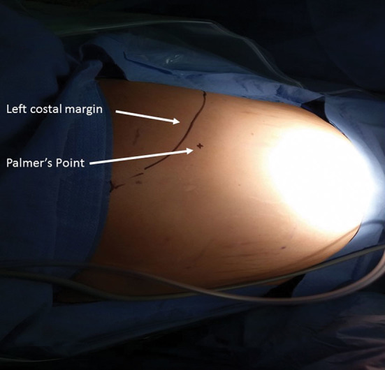
Palmer’s point is our preferred site for Veress needle insertion, lying two fingerbreadths below the left subcostal margin in the midclavicular line

The path of the Veress needle (arrow) is visualised using real time ultrasonography
Similar articles
-
Reply to safety of Veress needle insertion in laparoscopic bariatric surgery.Surg Laparosc Endosc Percutan Tech. 2014 Jun;24(3):283. doi: 10.1097/SLE.0000000000000069. Surg Laparosc Endosc Percutan Tech. 2014. PMID: 24887543 No abstract available.
-
Safe Veress needle insertion.J Hepatobiliary Pancreat Surg. 2006;13(3):225-7. doi: 10.1007/s00534-005-1024-x. J Hepatobiliary Pancreat Surg. 2006. PMID: 16708299
-
Safety of Veress needle insertion in laparoscopic bariatric surgery.Surg Laparosc Endosc Percutan Tech. 2014 Feb;24(1):e1-4. doi: 10.1097/SLE.0b013e31828f6cfd. Surg Laparosc Endosc Percutan Tech. 2014. PMID: 24487166
-
Laparoscopic needles and trocars: an overview of designs and complications.J Laparoendosc Surg. 1992 Apr;2(2):117-25. doi: 10.1089/lps.1992.2.117. J Laparoendosc Surg. 1992. PMID: 1534491 Review.
-
The role of optical access trocars in laparoscopic surgery.Surg Technol Int. 2005;14:61-7. Surg Technol Int. 2005. PMID: 16525956 Review.
Cited by
-
Computer-aided Veress needle guidance using endoscopic optical coherence tomography and convolutional neural networks.J Biophotonics. 2022 May;15(5):e202100347. doi: 10.1002/jbio.202100347. Epub 2022 Feb 11. J Biophotonics. 2022. PMID: 35103420 Free PMC article.
References
-
- Azevedo JL, Azevedo OC, Miyahira SA et al. . Injuries caused by Veress needle insertion for creation of pneumoperitoneum: a systematic literature review. Surg Endosc 2009; : 1,428–1,432. - PubMed
-
- Schäfer M, Lauper M, Krähenbühl L. Trocar and Veress needle injuries during laparoscopy. Surg Endosc 2001; : 275–280. - PubMed
-
- Jiang X, Anderson C, Schnatz PF. The safety of direct trocar versus Veress needle for laparoscopic entry: a meta-analysis of randomized clinical trials. J Laparoendosc Adv Surg Tech A 2012; : 362–370. - PubMed
MeSH terms
LinkOut - more resources
Full Text Sources
Other Literature Sources
Medical