. 2017 Oct 12;8(10):e3110.
doi: 10.1038/cddis.2017.523.

p38α MAPK antagonizing JNK to control the hepatic fat accumulation in pediatric patients onset intestinal failure

Affiliations

p38α MAPK antagonizing JNK to control the hepatic fat accumulation in pediatric patients onset intestinal failure

Yongtao Xiao et al. Cell Death Dis. .

Abstract

The p38α mitogen-activated protein kinase (MAPK) has been related to gluconeogenesis and lipid metabolism. However, the roles and related mechanisms of p38α MAPK in intestinal failure (IF)-associated liver steatosis remained poor understood. Here, our experimental evidence suggested that p38α MAPK significantly suppressed the fat accumulation in livers of IF patients mainly through two mechanisms. On the one hand, p38α MAPK increased hepatic bile acid (BA) synthesis by upregulating the expression of the rate-limiting enzyme cholesterol 7-α-hydroxylase (CYP7A1) and peroxisome proliferator-activated receptor γ coactivator-1α (PGC-1α), which in turn activated the transcription of the CYP7A1. On the other hand, p38α MAPK promoted fatty acid (FA) β-oxidation via upregulating peroxisome proliferator-activated receptor alpha (PPARα) and its transcriptional target genes carnitine palmitoyltransferase 1A (CPT1A) and peroxisomal acyl-coenzyme aoxidase 1 (ACOX1). Dual luciferase assays indicated that p38α MAPK increased the transcription of PPARα, PGC-1α and CYP7A1 by upregulating their promoters' activities. In addition, in vitro and in vivo assays indicated p38α MAPK negatively regulates the hepatic steatosis by controlling JNK activation. In conculsion, our findings demonstrate that hepatic p38α MAPK functions as a negative regulator of liver steatosis in maintaining BA synthesis and FAO by antagonizing the c-Jun N-terminal kinase (JNK).


Conflict of interest statement

The authors declare no conflict of interest.

Figures

Figure 1
Figure 1
Liver steatosis correlated with the duration of parenteral nutrition (PN) and related to the reducing bile acid (BA) contents in the liver tissues of in pediatric intestinal failure (IF) patients. (a) Representative images of haematoxylin and eosin (H&E) and transmission electron microscopy (TEM) in the liver tissues of IF patients with or without steatosis. (b,c) Quantification of the levels of hepatic triglyceride (TG) and phospholipids (PL) in IF patients. (d,e) The contents of hepatic TG and PL were positively correlated with PN duration. (f,g) The levels of BA in the liver and serum reduced in the patients with steatosis compared with the ones without steatosis. (h) The primary BA cholic acid (CA) and chenodeoxycholic acid (CDCA) decreased in the livers of patients with steatosis, related to the ones without steatosis. Scale bar=50 μm, 2 μm; Arrows indicate the fatty drops (a) with steatosis (n=6–10) and without steatosis (n=8–14) *P<0.05, **P<0.01
Figure 2
Figure 2
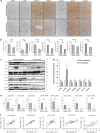
The p38α MAPK activation was decreased in livers of IF patients with steatosis and associated with expression of cholesterol 7-α-hydroxylase (CYP7A1), proliferator-activated receptor α coactivator-1 (PGC-1α) and nuclear receptor peroxisome proliferator-activated receptor α (PPARα). (a) Representative images of p-p38, p-JNK, CYP7A1, PGC-1α, PPARα, CPT1A and ACOX1 stainings in the liver tissues of IF patients without steatosis (n=14) and with steatosis (n=10). (b) Quantification of the results in panel A. (c) Western blot analyses of the p-p38, p-JNK, CYP7A1, PGC-1α, PPARα, CPT1A and ACOX1 protein levels in the livers of IF patients with or without steatosis. (d) Quantification of the protein results in panel C. (e) qRT- PCR analyses determine the levels of p38α, JNK1/2, CYP7A1, PGC-1α, PPARα, CPT1A and ACOX1 mRNAs in the IF patients. (f) The relationships between p38α mRNA and mRNA levels of CYP7A1, PGC-1α, PPARα, and between PGC-1α mRNA and CYP7A1 mRNA, PPARα mRNA in the liver tissues of the IF patients with Pearson’s correlations. Scale bar=25 μm *P<0.05, **P<0.01
Figure 3
Figure 3
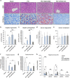
p38α MAPK and JNK have opposite effects on PN-associated hepatic steatosis. (a) H&E and Oil Red O staining showed that p38α MAPK inhibition with SB203580 treatment increased the fat accumulated in the liver of PN-fed rats. In contrast, JNK inhibition with SP600125 treatment suppressed the PN-associated steatosis. (b–e) The alteration of levels of the triglyceride (TG) and phospholipids (PL) in liver and serum from groups of Sham, PN, PN+SB203580 and PN+SP600125. (f,g) The levels of total BA in the liver and serum from groups of Sham, PN, PN+SB203580 and PN+SP600125. (h) The measurement of primary BA including cholic acid (CA), chenodeoxycholic acid (CDCA) and α,β,ω-muricholic acid (α-,β-,ωMCA) in livers from groups of Sham, PN, PN+SB203580 and PN+SP600125. Scale bar=25 μm *P<0.05, **P<0.01
Figure 4
Figure 4
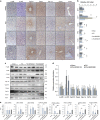
p38α MAPK opposing JNK to suppress the expression of CYP7A1, PGC-1α, PPARα, CPT1A and ACOX1 in vivo. (a) Representative images of p-p38, p-JNK, CYP7A1, PGC-1α, PPARα, CPT1A and ACOX1 stainings in the liver tissues of Sham, PN, PN+SB203580 and PN+SP600125 groups. (b) Quantification of the results in panel A. (c) Western blot analyses of the p-p38, p-JNK,CYP7A1, PGC-1α, PPARα, CPT1A and ACOX1 protein levels in the livers of Sham, PN, PN+SB203580 and PN+SP600125 groups. (d) Quantification of the protein results in panel C. (e) qRT-PCR analyses determine the levels of p38α, JNK1/2, CYP7A1, PGC-1α, PPARα, CPT1A and ACOX1 mRNAs in the livers of Sham, PN, PN+SB203580 and PN+SP600125 groups. Scale bar=25 μm *P<0.05, **P<0.01
Figure 5
Figure 5
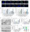
p38α MAPK antagonistically JNK to control the palmitate (PA)-mediated lipogenesis. (a) Representative images of neutral lipids staining in the indicated cells. (b,c) Quantification of the contents of hepatic triglyceride (TG) and phospholipids (PL) in indicated treatments. (d) Western blot analyses for the p-p38, p-JNK, CYP7A1, PGC-1α, PPARα, CPT1A and ACOX1 protein levels in the human primary hepatic cell with indicated treatments. (e–g) Relative promoter activity of CYP7A1, PGC-1α and PPARα with indicated treatments. (h) The scheme illustrates a potential mechanism by which p38α MAPK controls the fatty acid metabolism in the livers of IF patients. Scale bar=25 μm *P<0.05, **P<0.01

Similar articles

Cited by

References

    1. Wiles A, Woodward JM. Recent advances in the management of intestinal failure-associated liver disease. Curr Opin Clin Nutr Metab Care 2009; 12: 265–272. - PubMed
    1. Carter BA, Karpen SJ. Intestinal failure-associated liver disease: management and treatment strategies past, present, and future. Semin Liver Dis 2007; 27: 251–258. - PubMed
    1. Lauriti G, Zani A, Aufieri R, Cananzi M, Chiesa PL, Eaton S et al. Incidence, prevention, and treatment of parenteral nutrition-associated cholestasis and intestinal failure-associated liver disease in infants and children: a systematic review. JPEN 2014; 38: 70–85. - PubMed
    1. Kurvinen A, Nissinen MJ, Andersson S, Korhonen P, Ruuska T, Taimisto M et al. Parenteral plant sterols and intestinal failure-associated liver disease in neonates. J Pediatr Gastroenterol Nutr 2012; 54: 803–811. - PubMed
    1. Nehra D, Fallon EM, Puder M. The prevention and treatment of intestinal failure-associated liver disease in neonates and children. Surg Clin N Am 2011; 91: 543–563. - PubMed

MeSH terms

Substances