Review
. 2017 Oct 12;9(10):314.
doi: 10.3390/toxins9100314.

Plant Ribosome-Inactivating Proteins: Progesses, Challenges and Biotechnological Applications (and a Few Digressions)

Affiliations
Review

Plant Ribosome-Inactivating Proteins: Progesses, Challenges and Biotechnological Applications (and a Few Digressions)

Maria Serena Fabbrini et al. Toxins (Basel). .

Abstract

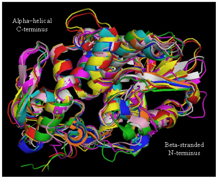
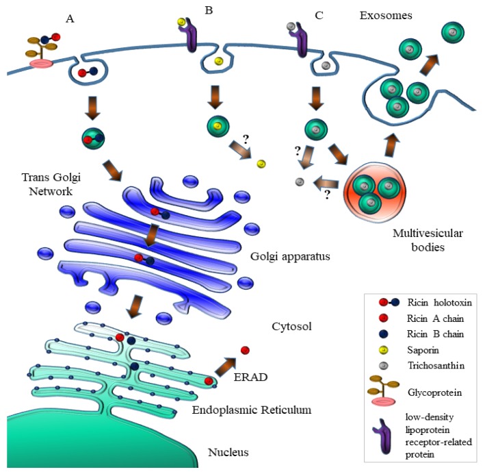
Plant ribosome-inactivating protein (RIP) toxins are EC3.2.2.22 N-glycosidases, found among most plant species encoded as small gene families, distributed in several tissues being endowed with defensive functions against fungal or viral infections. The two main plant RIP classes include type I (monomeric) and type II (dimeric) as the prototype ricin holotoxin from Ricinus communis that is composed of a catalytic active A chain linked via a disulphide bridge to a B-lectin domain that mediates efficient endocytosis in eukaryotic cells. Plant RIPs can recognize a universally conserved stem-loop, known as the α-sarcin/ ricin loop or SRL structure in 23S/25S/28S rRNA. By depurinating a single adenine (A4324 in 28S rat rRNA), they can irreversibly arrest protein translation and trigger cell death in the intoxicated mammalian cell. Besides their useful application as potential weapons against infected/tumor cells, ricin was also used in bio-terroristic attacks and, as such, constitutes a major concern. In this review, we aim to summarize past studies and more recent progresses made studying plant RIPs and discuss successful approaches that might help overcoming some of the bottlenecks encountered during the development of their biomedical applications.

Keywords: ER-stress; nanovectors; plant ribosome inactivating proteins; saporin; targeted drug delivery.


Conflict of interest statement

The authors declare no conflict of interest.

Figures

Figure 1
Figure 1
Amino acid sequence alignment of different type I RIPs compared to ricin (RTA), abrin and cinnamomin catalytic A chains by T-Coffee. Color shades indicate levels of amino acid homology between the aligned sequences. Conserved amino acids are identified with an asterisk and residues crucial for the catalytic activity are arrowed: Tyr72, Tyr120, Glu176, Arg179 and Trp208 in the sequence of saporin.
Figure 2
Figure 2
Three-dimensional reconstruction of catalytic cleft of saporin obtained by Swiss PDB Viewer (v4.0.4, SIB—Swiss Institute of Bioinformatics, Lausanne, Switzerland). Conserved residues crucial for the RIP signature are colored: Tyr72 (yellow), Tyr120 (red), Glu176 (orange), Arg179 (green) and Trp208 (blue). Hydrogen bonds among key residues are shown in orange.
Figure 3
Figure 3
Three-dimensional structure of different type I RIPs and ricin A chain (RTA). Superimposition of secondary structure elements of Saporin (red, PDB code 1QI7), Gelonin (pink, 3KU0), PAP (magenta, 1GIK), Trichosanthin (cyan, 1QD2), Dianthin (yellow, 1RL0), Bouganin (grey, 3CTK), Momordin (orange, 1 MOM), Momorcharin (blue, 1AHA), RTA (green, 1J1 M), modified from [31].
Figure 4
Figure 4
Kyte–Doolittle hydrophobicity Plot of the signal peptides of type I RIPs, tobacco BiP and PrP protein obtained by BioEdit software (version 7.0.9.0, Ibis Therapeutics, Carlsbad, CA, USA). Sequences used are shown under the plots.
Figure 5
Figure 5
Phylogenetic tree of type I and type II RIPs obtained using the amino acidic sequences aligned with the Clustal Omega on-line software (EMBL-EBI, Hinxton, UK) with standard parameters. The branch length represents the number of changes that occurred in that branch.
Figure 6
Figure 6
Schematic representation of the intoxication routes followed by ricin (A); saporin (B) and trichosanthin (C), modified from [56].
Figure 7
Figure 7
Present and future applications of RIPs. A schematic representation summarizes some of the potential application of a toxic plant RIP (green star and 3D-ribbon) starting with a classical Immunotoxin made by IgG chemically conjugated to native RIPs, which may be further transformed (left) into a fusion recombinant chimera or (right) into single chain variable fusions to RIP or even simply to DNA constructs encoding the plant RIP with all these different molecules being deliverable via targeted exosomes or engineered nanoparticles even for diagnostic purposes with MnFe2O4 nanoparticles (see text below).

References

    1. Stirpe F. Ribosome-inactivating proteins: From toxins to useful proteins. Toxicon: Off. J. Int. Soc. Toxinol. 2013;67:12–16. doi: 10.1016/j.toxicon.2013.02.005. - DOI - PubMed
    1. Endo Y., Tsurugi K. RNA N-glycosidase activity of ricin A-chain. Mechanism of action of the toxic lectin ricin on eukaryotic ribosomes. J. Biol. Chem. 1987;262:8128–8130. - PubMed
    1. Endo Y., Mitsui K., Motizuki M., Tsurugi K. The mechanism of action of ricin and related toxic lectins on eukaryotic ribosomes. The site and the characteristics of the modification in 28 S ribosomal RNA caused by the toxins. J. Biol. Chem. 1987;262:5908–5912. - PubMed
    1. Benatti L., Saccardo M.B., Dani M., Nitti G., Sassano M., Lorenzetti R., Lappi D.A., Soria M. Nucleotide sequence of cDNA coding for saporin-6, a type-1 ribosome-inactivating protein from Saponaria officinalis. Eur. J. Biochem. 1989;183:465–470. doi: 10.1111/j.1432-1033.1989.tb14951.x. - DOI - PubMed
    1. Arabidopsis Genome I. Analysis of the genome sequence of the flowering plant Arabidopsis thaliana. Nature. 2000;408:796–815. doi: 10.1038/35048692. - DOI - PubMed

Publication types

Substances