. 2017 Oct 12;15(10):e2003790.
doi: 10.1371/journal.pbio.2003790. eCollection 2017 Oct.

The sea cucumber genome provides insights into morphological evolution and visceral regeneration

Affiliations

The sea cucumber genome provides insights into morphological evolution and visceral regeneration

Xiaojun Zhang et al. PLoS Biol. .

Abstract

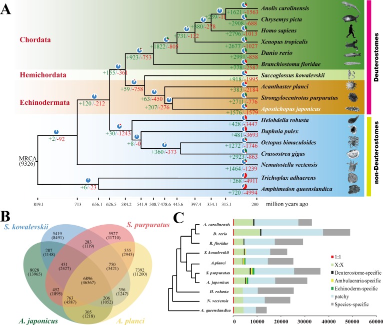
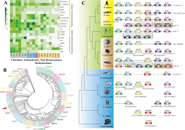
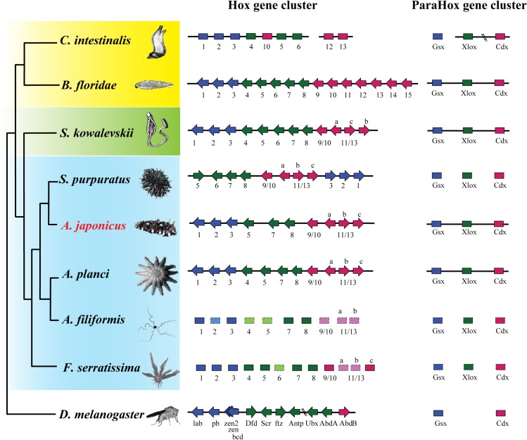
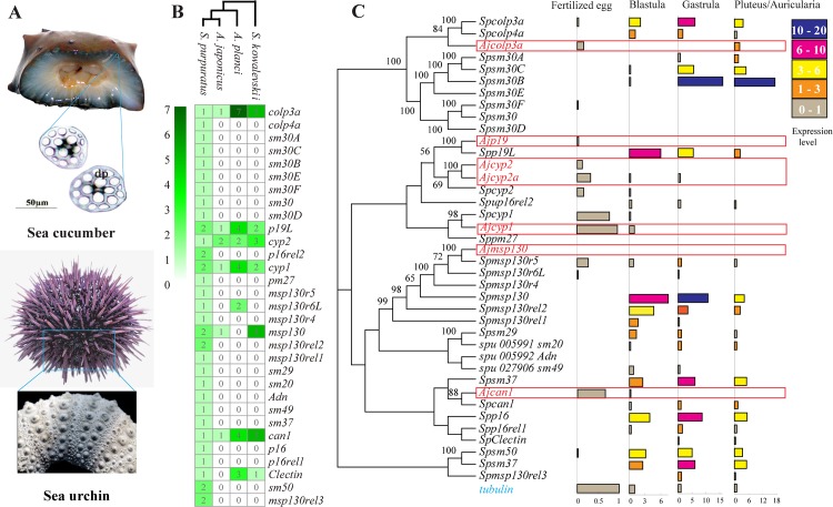
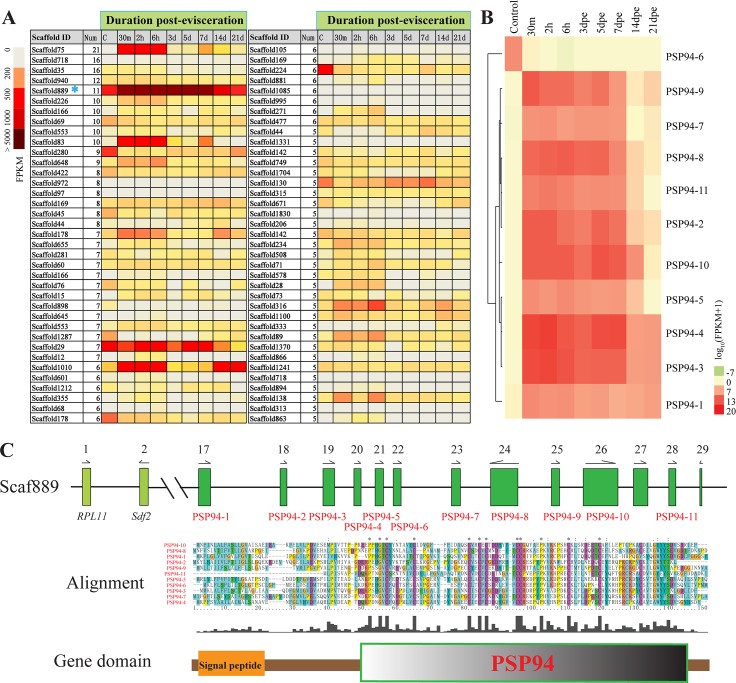
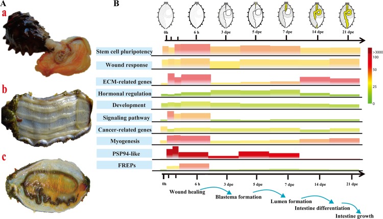
Apart from sharing common ancestry with chordates, sea cucumbers exhibit a unique morphology and exceptional regenerative capacity. Here we present the complete genome sequence of an economically important sea cucumber, A. japonicus, generated using Illumina and PacBio platforms, to achieve an assembly of approximately 805 Mb (contig N50 of 190 Kb and scaffold N50 of 486 Kb), with 30,350 protein-coding genes and high continuity. We used this resource to explore key genetic mechanisms behind the unique biological characters of sea cucumbers. Phylogenetic and comparative genomic analyses revealed the presence of marker genes associated with notochord and gill slits, suggesting that these chordate features were present in ancestral echinoderms. The unique shape and weak mineralization of the sea cucumber adult body were also preliminarily explained by the contraction of biomineralization genes. Genome, transcriptome, and proteome analyses of organ regrowth after induced evisceration provided insight into the molecular underpinnings of visceral regeneration, including a specific tandem-duplicated prostatic secretory protein of 94 amino acids (PSP94)-like gene family and a significantly expanded fibrinogen-related protein (FREP) gene family. This high-quality genome resource will provide a useful framework for future research into biological processes and evolution in deuterostomes, including remarkable regenerative abilities that could have medical applications. Moreover, the multiomics data will be of prime value for commercial sea cucumber breeding programs.


Conflict of interest statement

The authors have declared that no competing interests exist.

Figures

Fig 1
Fig 1. A schematic representation of the genomic characteristics of A. japonicus.
Track 1: 22 linkage groups of the genome. Track 2: anchored scaffolds to each linkage group. Long bars represent scaffolds with a length > 500 Kb; short bars represent scaffolds with a length ≤ 500 Kb. Track 3: protein-coding genes located on scaffolds. Red stands for genes on the forward strand, and green stands for genes on the reverse strand. Track 4: distributions of 5 significantly expanded gene families in the genome. The 5 gene families are fibrinogen-related proteins (FREPs) (F), retrovirus-related Pol polyprotein from transposon (R), nucleotide-binding oligomerization domain-like receptors (NLR) family caspase recruitment domain (CARD) domain-containing protein (N), tyrosine-protein kinase receptor (T), and zinc finger CysCysHisCys (CCHC) domain-containing protein (Z). These expanded gene families show a clustered distribution in the genome. FREPs mainly clustered at LG02, and NLR chiefly accumulated at LG04. Track 5: distribution of microRNA (miRNA) in the genome. Most miRNAs are distributed in clusters across the genome (LG01, LG10, LG11, LG12, and LG22). The data underlying Fig 1 can be found in S3 Data.
Fig 2
Fig 2. Comparative genomic analysis between A. japonicus and other metazoans.
(A) Phylogenetic placement of A. japonicus within the metazoan tree. The numbers on the branches indicate the number of gene gains (+) and the number of gene losses (−), which are also displayed as bar plots: gene gain (in green), gene loss (in red), and the remaining gene families (in blue). The divergence times were estimated and displayed below the phylogenetic tree. Image credits: Robert Michniewicz; Kobie Mercury-Clarke; Vector Open Stock, Patrick Narbonne, David E. Simpson, John B. Gurdon; Lars Simonsen; Freshwater and Marine Image Bank; Encyclopædia Britannica; public domain; Jerry Kirkhart; authors' own; Johny Ha; Virginia Gewin; public domain; public domain; Cnidaria; Michael Eitel, Hans-Jürgen Osigus, Rob DeSalle, Bernd Schierwater; Maja Adamska. (B) The shared and unique gene families in 4 species of Ambulacraria are shown in the Venn diagram. There are 763 gene families shared by 3 echinoderms. (C) Comparison of the gene repertoire of 10 metazoan genomes. Here, "1:1" indicates single-copy genes; "X:X" indicates orthologous genes present in multiple copies in all the 9 species, where X means 1 or more orthologs per species; and "patchy" indicates the existence of other orthologs that are present in at least 1 genome. A. japonicus and S. purpuratus show a similar distribution of gene repertoire. The data underlying Fig 2 can be found in S3 Data.
Fig 3
Fig 3. Gene families and gene clusters related to notochord and pharyngeal gill slit formation.
(A) Distribution pattern of genes related to notochord and stomochord formation in metazoans. These genes are all present in deuterostomes, but some of them are present in nondeuterostomes. (B) Phylogenetic analysis of the fibroblast growth factor (FGF) gene family in metazoans. Different genes of the FGF gene family are distinguished with different colors. The FGF gene family is important for transcriptional activation of bruchury in notochord formation, but only 1 such gene exists in the genome of A. japonicus (red arrow) and the other 2 echinoderms (yellow background), S. purpuratus (Spur_15908_340) and A. planci (Apla_2356_405). The data underlying Fig 3A and 3B can be found in S3 Data. (C) A gene cluster related to the pharyngeal gill slit. The genes connected on a line indicate that they are clustered in order in the genome. This gene cluster is conserved among deuterostomes, whereas it is incomplete and shows poor synteny within nondeuterostomes. Image credits: Robert Michniewicz; Kobie Mercury-Clarke; Patrick Narbonne, David E. Simpson, John B. Gurdon; Vector Open Stock; Lars Simonsen; Freshwater and Marine Image Bank; Encyclopædia Britannica; public domain; Jerry Kirkhart; authors' own; Martin Cooper; public domain; public domain; Cnidaria; Freshwater and Marine Image Bank; Maja Adamska; Johny Ha; Virginia Gewin; Michael Eitel, Hans-Jürgen Osigus, Rob DeSalle, Bernd Schierwater.
Fig 4
Fig 4. The conserved homeobox gene clusters in the genome of A. japonicus.
A. planci, Amphiura filiformis, and Florometra serratissima belong to Echinodermata class Asteroidea, Ophiuroidea, and Crinoidea, respectively. The general phylogenetic tree shows the Hox and ParaHox cluster order and genes. Hox4 and Hox6 are not found in the genome of A. japonicus. Different colors indicate different Hox gene groups, including anterior (blue), central (green), and posterior (red). The arrow indicates the direction of transcription. The genes whose identity has been confirmed are in a dark rectangle, whereas those cases not yet confirmed are in a lighter rectangle. The Hox cluster images of Branchiostoma floridae, S. purpuratus, A. filiformis, and F. serratissima were modified from Byrne et al. 2016 [34]. Image credits: "Introduction to zoology; a guide to the study of animals, for the use of secondary schools" (1900) Macmillan; Freshwater and Marine Image Bank; Encyclopædia Britannica; Jerry Kirkhart; authors' own; public domain; Freshwater and Marine Image Bank; National Oceanic and Atmospheric Administration; Martin Cooper.
Fig 5
Fig 5. Gene families related to skeletogenesis and biomineralization in the genome of A. japonicus.
(A) Comparison of the skeleton from A. japonicus and S. purpuratus. (B) The comparison of the copy numbers of biomineralization-related genes in 3 echinoderms, A. japonicus, S. purpuratus, and A. planci, and the hemichordate S. kowalevskii. Biomineralization-related genes are apparently contracted in A. japonicus. (C) The expression levels of biomineralization-related genes at early developmental stages of A. japonicus (red) and S. purpuratus (black). The expression level of these genes at different development stages is calculated relative to a housekeeping gene (tubulin). Compared with sea urchin, there is a significant gene loss in the A. japonicus genome, and the expression levels for those genes at different development stages are relatively low. The data underlying Fig 5B and 5C can be found in S3 Data.
Fig 6
Fig 6. Prostatic secretory protein of 94 amino acids (PSP94)-like gene family in the genome of A. japonicus.
(A) A heatmap showing the expression profile of tandem-duplicated gene clusters during the intestinal regeneration of A. japonicus. It depicts the cluster ID, the repeat number, and the expression profile. The color represents the relative expression level (fragments per kilobase of transcript per million mapped reads [FPKM] value) of the gene clusters during the intestinal regeneration. The Scaffold889 with 11 tandem-duplicated genes (blue asterisk) shows significant up-regulation post evisceration. (B) The detailed expression of 11 PSP94-like genes located on Scaffold889 at different time points post evisceration. The scale covers log expression values. Genes are clustered by Euclidean distance of the log10(FPKM+1) value and grouped with average-linkage clustering. (C) The distribution and alignment of the PSP94-like gene family. Eleven PSP94-like genes are tandemly arranged on Scaffold889. Arrows denote the transcriptional orientation. The amino acid sequence alignment and domain prediction of proteins coded by these PSP94-like genes show highly conserved cysteine residues in the PSP94 domain. The data underlying Fig 6B and 6C can be found in S3 Data.
Fig 7
Fig 7. Genes involved in the intestine regeneration of A. japonicus.
(A) Regeneration diagram showing A. japonicus (a) undergoing evisceration, (b) immediately post evisceration, and (c) after complete recovery. (B) A heatmap showing the expression profile of molecular events applicable to the intestinal regeneration of A. japonicus. It depicts the molecular events identified in this study in relation to the morphological diversification of regeneration in a diagrammatic sketch. The height and the color of trapezium represent the relative expression level (fragments per kilobase of transcript per million mapped reads [FPKM] value per gene) during the regeneration. High-expressed and low-expressed genes are labeled in red and green, respectively. Extracellular matrix (ECM)-related genes (collagen and fibropellin), signaling pathways (Wnt, bone morphogenetic protein-related genes, and epidermal growth factor-related genes), and myogenesis-related genes (tubulin) are activated at the early stage of regeneration (0–3 days post evisceration [dpe]). During the middle stage of regeneration (3–7 dpe), factors related to hormonal regulation are up-regulated. In the late stages of regeneration, ECM-related genes (tenascin, FRAS1, and collagen) and myogenesis-related genes (actin and myosin) show up-regulated expression. The physiological events happening during regeneration are shown below the x-axis. The data underlying Fig 7B can be found in S3 Data. FREP, fibrinogen-related protein.

References

    1. Sodergren E, Weinstock GM, Davidson EH, Cameron RA, Gibbs RA, Angerer RC, et al. The genome of the sea urchin Strongylocentrotus purpuratus. Science. 2006;314: 941–952. doi: 10.1126/science.1133609 - DOI - PMC - PubMed
    1. Hall MR, Kocot KM, Baughman KW, Fernandez-Valverde SL, Gauthier MEA, Hatleberg WL, et al. The crown-of-thorns starfish genome as a guide for biocontrol of this coral reef pest. Nature. 2017;544(7649): 231–234. doi: 10.1038/nature22033 - DOI - PubMed
    1. Rychel AL, Swalla BJ. Regeneration in hemichordates and echinoderms. Stem Cells in Mar Organ. 2009: 245–265.
    1. Carnevali MC. Regeneration in Echinoderms: repair, regrowth, cloning. ISJ-Invertebr Surviv J. 2006;3: 64–76.
    1. Mashanov VS, García-Arrarás JE. Gut regeneration in holothurians: a snapshot of recent developments. Biol Bull. 2011;221: 93–109. doi: 10.1086/BBLv221n1p93 - DOI - PubMed