Comparative Study
. 2018 Jan;232(1):134-145.
doi: 10.1111/joa.12707. Epub 2017 Oct 10.

Anatomy and development of the extrahepatic biliary system in mouse and rat: a perspective on the evolutionary loss of the gallbladder

Affiliations
Comparative Study

Anatomy and development of the extrahepatic biliary system in mouse and rat: a perspective on the evolutionary loss of the gallbladder

Hiroki Higashiyama et al. J Anat. 2018 Jan.

Abstract

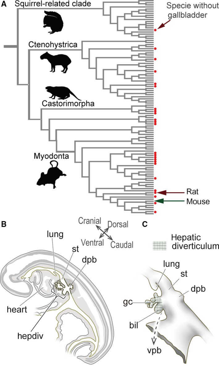
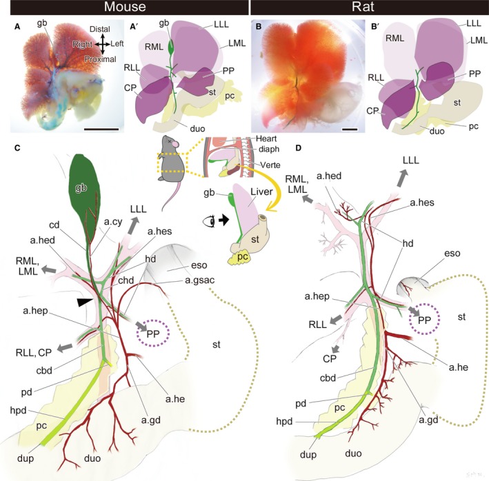
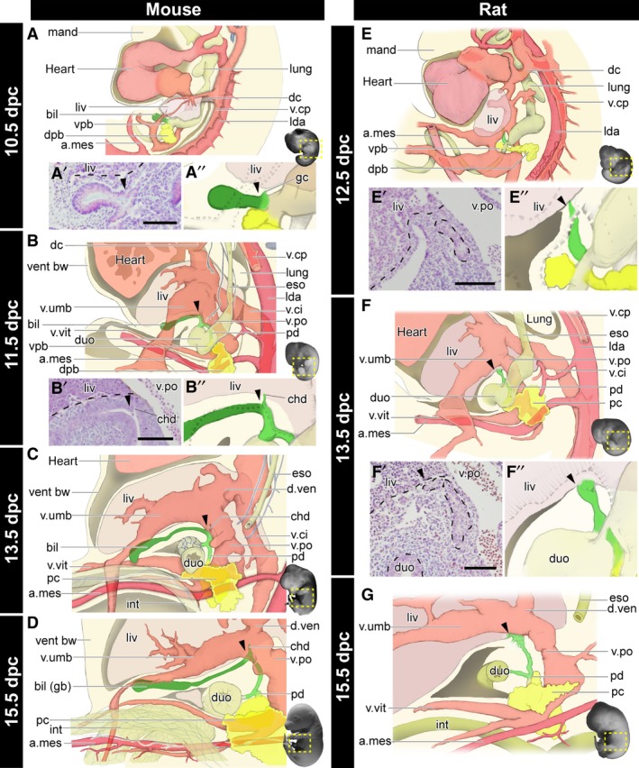
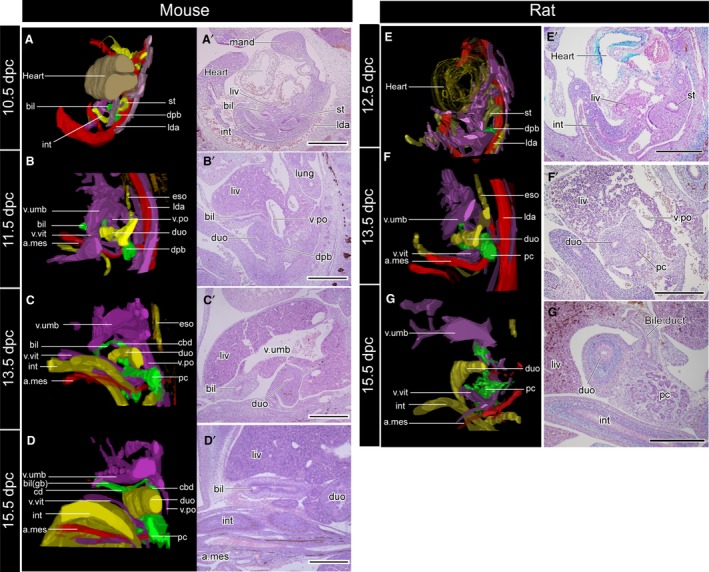
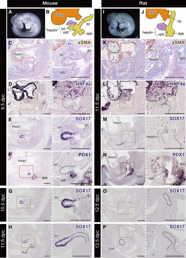
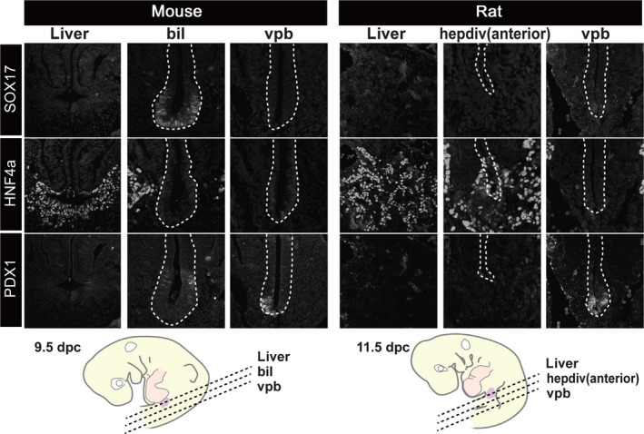
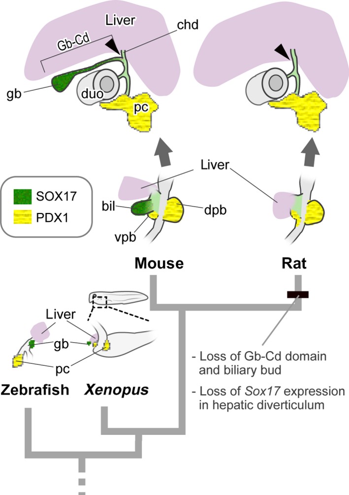
The gallbladder is the hepatobiliary organ for storing and secreting bile fluid, and is a synapomorphy of extant vertebrates. However, this organ has been frequently lost in several lineages of birds and mammals, including rodents. Although it is known as the traditional problem, the differences in development between animals with and without gallbladders are not well understood. To address this research gap, we compared the anatomy and development of the hepatobiliary systems in mice (gallbladder is present) and rats (gallbladder is absent). Anatomically, almost all parts of the hepatobiliary system of rats are topographically the same as those of mice, but rats have lost the gallbladder and cystic duct completely. During morphogenesis, the gallbladder-cystic duct domain (Gb-Cd domain) and its primordium, the biliary bud, do not develop in the rat. In the early stages, SOX17, a master regulator of gallbladder formation, is positive in the murine biliary bud epithelium, as seen in other vertebrates with a gallbladder, but there is no SOX17-positive domain in the rat hepatobiliary primordia. These findings suggest that the evolutionary loss of the Gb-Cd domain should be translated simply as the absence of a biliary bud at an early stage, which may correlate with alterations in regulatory genes, such as Sox17, in the rat. A SOX17-positive biliary bud is clearly definable as a developmental module that may be involved in the frequent loss of gallbladder in mammals.

Keywords: biliary tract; gallbladder; modularity; mouse; rat.


Figures

Figure 1
Figure 1
Diversity in the presence/absence of a gallbladder in rodents, and the definition of the mammalian hepatobiliary primordia. (A) Gallbladder evolution in rodents. The phylogenetic framework is based on Fabre et al. (2012), and the topology of the tree was arranged in Mesquite (Maddison & Maddison, 2011). The presence of a gallbladder is from Gorham & Ivy (1938) and Nzalak et al. (2010). The red dots indicate species that do not have gallbladders. The silhouette images are from PhyloPic (http://phylopic.org/). For more details, see Fig. S1. (B) Scheme of the left lateral view of the sagittal section of a pig embryo at the pharyngula stage. The hepatobiliary primordia arises from the foregut at the level of the septum transversum, caudal to the heart. (C) The hepatobiliary primordia in a pig embryo. The meshed domain is identified as the ‘hepatic diverticulum’ (sensu Patten, 1927). The diverticulum differentiates into the primordial liver, and biliary and ventral pancreatic buds. Of these, the biliary bud provides most of the extrahepatic biliary tract. The figures in (B) and (C) are redrawn from Patten (1927). bil, biliary bud; dpb, dorsal pancreatic bud; gc, glandular cord; hepdiv, hepatic diverticulum; lung, lung bud; st, stomach; vpb, ventral pancreatic bud. [Correction added on 24 October 2017, after first online publication: the abbreviations cited on this figure was added on figure caption]
Figure 2
Figure 2
Gross anatomy of the extrahepatic biliary system in the mouse and rat. (A,A′) The morphology of the whole hepatobiliary system of the mouse. (B,B′) The morphology of the whole hepatobiliary system of the rat. (C,D) Anatomy of the hepatobiliary system of the mouse (C) and rat (D). The gallbladder and cystic duct are coloured in dark green, the hepatopancreatic duct is light green, and the other bile ducts are middle green. The arrowhead indicates the branching point of the cystic duct and the common hepatic duct. The grey arrows indicate the directions of the portal veins that supply the liver lobes. a.cy, arteria cystica; a.gd, arteria gastroduodenalis; a.gsac, arteria gastrica sinistra accessoria; a.he, arteria hepatica communis; a.hed, arteria hepatica dextra; a.hep, arteria hepatica propria; a.hes, arteria hepatica sinistra; cbd, common bile duct; cd, cystic duct; chd, common hepatic duct; CP, caudate process of the caudate lobe; diaph, diaphragm; duo, duodenum; dup, duodenal papilla; eso, oesophagus; gb, gallbladder; hd, hepatic duct; hpd, hepatopancreatic duct; LLL, left lateral lobe; LML, left medial lobe; pc, pancreas; pd, pancreatic duct; PP, papillary process of the caudate lobe; RLL, right lateral lobe; RML, right medial lobe; st, stomach; verte, vertebrae. Scale bar: 1 cm. [Correction added on 24 October 2017, after first online publication: the abbreviations cited on this figure was added on figure caption]
Figure 3
Figure 3
Morphogenesis of the biliary system in the mouse and rat. All panels are left lateral views. See also Figs 4 and S2 for the histological sections, digitally reconstructed models and latex‐injected models. (A–D) Developmental scheme of the hepatobiliary system in mice. The biliary bud (green), pancreas (yellow), artery (magenta), and vein (pink) are drawn. The gallbladder–cystic duct (Gb–Cd) domain (dark green) can be identified as the distal domain from the branching point (arrowheads) of the common hepatic duct (chd). Details of the common hepatic ducts are shown in panels (A’), (A’’) and (B’), (B’’). (E–G) The developmental scheme in rats. The arrowheads indicate the landmarks comparable with mice. The Gb–Cd domain is clearly absent. a.mes, arteria mesenterica cranialis; bil, biliary bud; chd, common hepatic duct; dc, ductus cuvieri; dpb, dorsal pancreatic bud; duo, duodenum; d.ven, ductus venosus; eso, oesophagus; gb, gallbladder; gc, glandular cord; int, intestine; lda, left dorsal aorta; liv, liver or liver primordium; lung, lung bud; mand, mandibular process; pc, pancreas; pd, pancreatic duct; v.ci, vena cava inferior; v.cp, vena cava posterior; vent bw, ventral bodywall; vpb, ventral pancreatic bud; v.po, vena portae; v.umb, vena umbilcailis; v.vit, vena vitelline. Scale bar: 500 μm. [Correction added on 24 October 2017, after first online publication: the abbreviations cited on this figure was added on figure caption]
Figure 4
Figure 4
Three‐dimensional reconstructed models (A–D) and the underlying histological sections (A′–D′). The topographical relationships of all samples are the same as in Fig. 3. The sections were subjected to immunohistochemical (IHC) staining for acetylated tubulin to visualise the peripheral nerves. The biliary tract is innervated by the intestinal part of the vagus, but the nerve supplies had not been formed at the developmental stages examined in the present study. a.mes, arteria mesenterica cranialis; bil, biliary bud; cbd, common bile duct; cd, cystic duct; dpb, dorsal pancreatic bud; duo, duodenum; eso, oesophagus; gb, gallbladder; int, intestine; lda, left dorsal aorta; liv, liver or liver primordium; lung, lung bud; mand, mandibular process; pc, pancreas; v.po, vena portae; v.umb, vena umbilcailis; v.vit, vena vitelline. Scale bar: 500 μm. [Correction added on 24 October 2017, after first online publication: the abbreviations cited on this figure was added on figure caption]
Figure 5
Figure 5
Molecular patterning of the hepatobiliary primordia. All panels are left lateral views. (A) The mouse embryo at 9.5 dpc. The scheme of the hepatobiliary primordia is shown in (B). ‘hepdiv’ in (B) indicates the presumptive domain that corresponds to the hepatic diverticulum of the 11.5 dpc rat in (J). (C–F) Histological sections of the same 9.5‐dpc embryo with immunohistochemical (IHC) staining for aSMA, HNF4a, SOX17 and PDX1. (G,H) Sections of 10.5‐ and 11.5‐dpc mice stained for SOX17. The SOX17‐positive domain is localised in the distal portion of the biliary bud. (I) A rat embryo at 11.5 dpc. The scheme of the hepatobiliary primordia is shown in (J). (K–N) Sections of the same 11.5‐dpc embryo with IHC staining in the same manner as the mouse. (M,N) Sections of 12.5‐ and 13.5‐dpc rats stained for SOX17. The black dotted line in the higher‐magnification panels indicates the extrahepatic biliary tract. bil, biliary bud; dc, ductus cuvieri; dpb, dorsal pancreatic bud; duo, duodenum; gc, glandular cord; hepdiv, hepatic diverticulum; liv, liver or liver primordium; vpb, ventral pancreatic bud. Scale bars: 200 μm. [Correction added on 24 October 2017, after first online publication: the abbreviations cited on this figure was added on figure caption].
Figure 6
Figure 6
Patterns of molecular markers that are common in the hepatobiliary primordia. Transverse sections were made at the levels shown in the scheme. The SOX17‐positive domain was clearly localised in the distal portion of the biliary bud in the mouse, but there was no equivalent signal in the rat embryo. HNF4a (liver tissue) and PDX1 were positive in the liver and pancreatic primordia in both animals. bil, biliary bud; hepdiv, hepatic diverticulum; vpb, ventral pancreatic bud. cale bars: 200 μm. [Correction added on 24 October 2017, after first online publication: the abbreviations cited on this figure was added on figure caption]
Figure 7
Figure 7
Evolutionary scenario of gallbladder loss in the rat. The schemes for zebrafish and Xenopus are drawn after Shin et al. (2012) and Zorn & Mason (2001), respectively. An obvious hepatic diverticulum is found in the ventral foregut in the rat, as seen in many other vertebrates. However, the SOX17‐positive biliary bud is completely lost in rats, while Sox17 is expressed in other animals that have gallbladders. bil, biliary bud; cd, cystic duct; chd, common hepatic duct; dpb, dorsal pancreatic bud; duo, duodenum; gb, gallbladder; pc, pancreas; vpb, ventral pancreatic bud. [Correction added on 24 October 2017, after first online publication: the abbreviations cited on this figure was added on figure caption]

References

    1. Berthoud HR, Kressel M, Neuhuber W (1992) An anterograde tracing study of the vagal innervation of rat liver, portal vein and biliary system. Anat Embryol 186, 431–442. - PubMed
    1. Biemar F, Argenton F, Schmidtke R, et al. (2001) Pancreas development in zebrafish: early dispersed appearance of endocrine hormone expressing cells and their convergence to form the definitive islet. Dev Biol 230, 189–203. - PubMed
    1. Breazile JE, Brown EM (1976) Anatomy In: The Biology of the Guinea Pig. (eds Wagner JE, Manning PJ.), pp. 53–62. New York, NY: Academic Press.
    1. Cardinale V, Wang Y, Carpino G, et al. (2011) Multipotent stem/progenitor cells in human biliary tree give rise to hepatocytes, cholangiocytes, and pancreatic islets. Hepatology 54, 2159–2172. - PubMed
    1. Carpino G, Cardinale V, Onori P, et al. (2012) Biliary tree stem/progenitor cells in glands of extrahepatic and intraheptic bile ducts: an anatomical in situ study yielding evidence of maturational lineages. J Anat 220, 186–199. - PMC - PubMed

Publication types

LinkOut - more resources