. 2018 Feb;33(2):283-297.
doi: 10.1002/jbmr.3305. Epub 2017 Nov 2.

Loss of Dnmt3b in Chondrocytes Leads to Delayed Endochondral Ossification and Fracture Repair

Affiliations

Loss of Dnmt3b in Chondrocytes Leads to Delayed Endochondral Ossification and Fracture Repair

Cuicui Wang et al. J Bone Miner Res. 2018 Feb.

Abstract

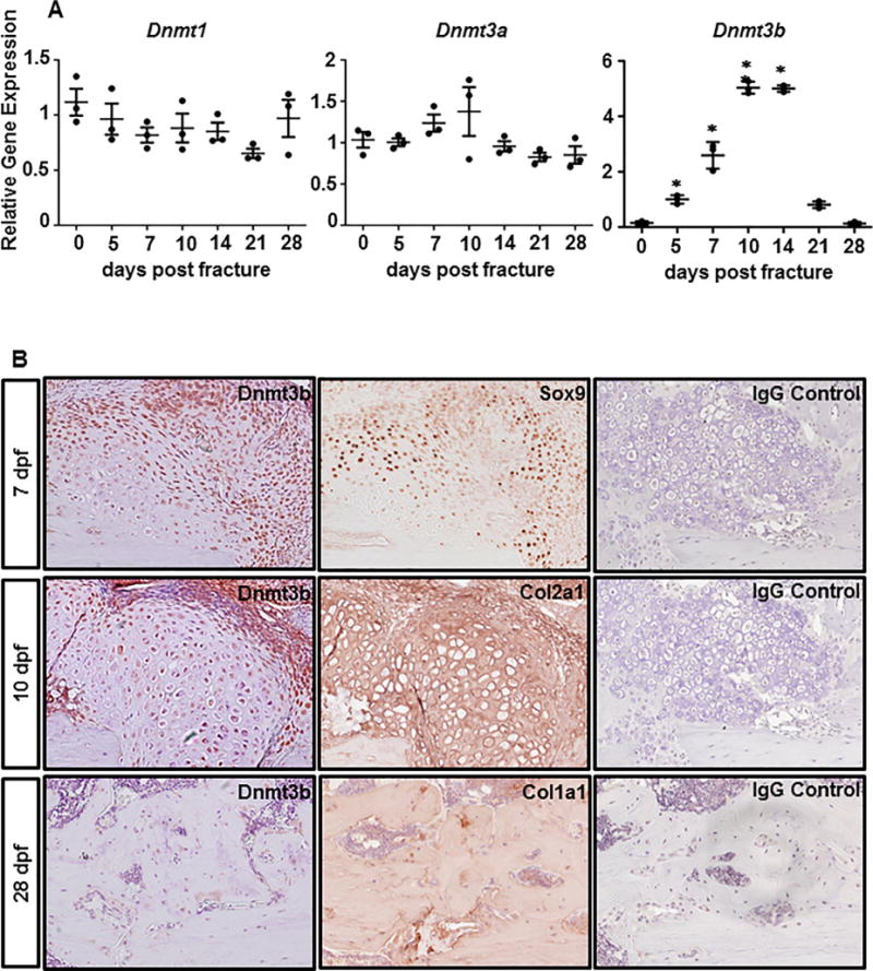
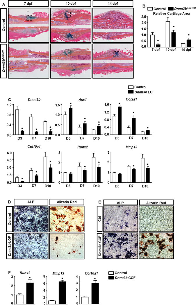
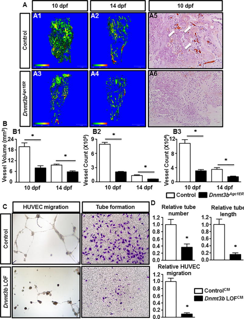
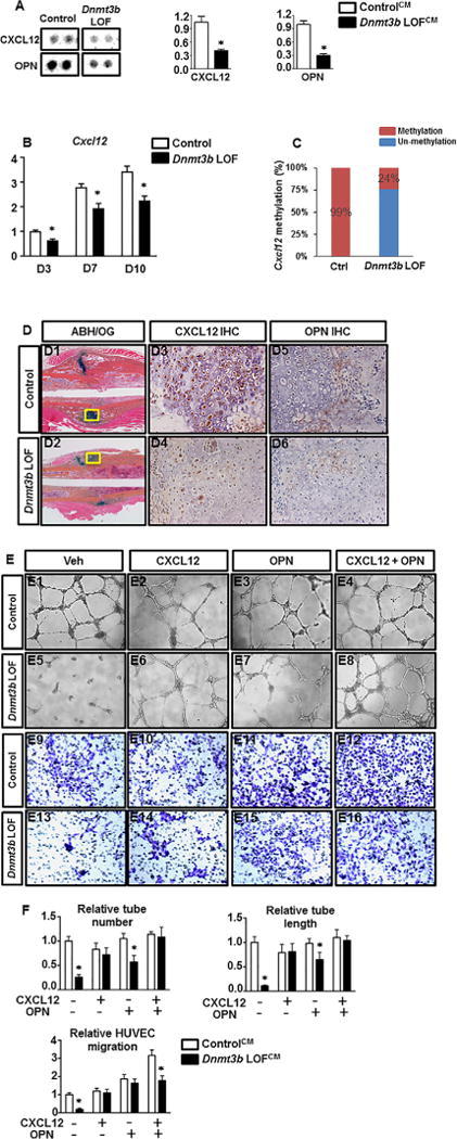
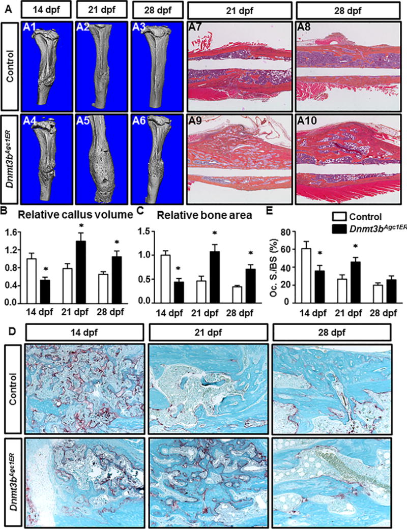
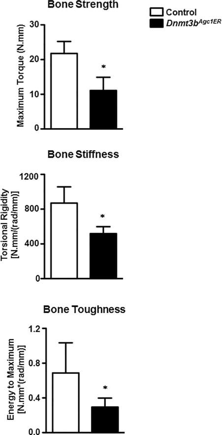
Despite advanced understanding of signaling mediated by local and systemic factors, the role of epigenetic factors in the regulation of bone regeneration remains vague. The DNA methyltransferases (Dnmts) Dnmt3a and Dnmt3b have tissue specific expression patterns and create unique methylation signatures to regulate gene expression. Using a stabilized murine tibia fracture model we find that Dnmt3b is induced early in fracture healing, peaks at 10 days post fracture (dpf), and declines to nearly undetectable levels by 28 dpf. Dnmt3b expression was cell-specific and stage-specific. High levels were observed in chondrogenic lineage cells within the fracture callus. To determine the role of Dnmt3b in fracture healing, Agc1CreERT2 ;Dnmt3bf/f (Dnmt3bAgc1ER ) mice were generated to delete Dnmt3b in chondrogenic cells. Dnmt3bAgc1ER fracture displayed chondrogenesis and chondrocyte maturation defect, and a delay in the later events of angiogenesis, ossification, and bone remodeling. Biomechanical studies demonstrated markedly reduced strength in Dnmt3bAgc1ER fractures and confirmed the delay in repair. The angiogenic response was reduced in both vessel number and volume at 10 and 14 dpf in Dnmt3bAgc1ER mice. Immunohistochemistry showed decreased CD31 expression, consistent with the reduced angiogenesis. Finally, in vitro angiogenesis assays with human umbilical vein endothelial cells (HUVECs) revealed that loss of Dnmt3b in chondrocytes significantly reduced tube formation and endothelial migration. To identify specific angiogenic factors involved in the decreased callus vascularization, a protein array was performed using conditioned media isolated from control and Dnmt3b loss-of-function chondrocytes. Several angiogenic factors, including CXCL12 and osteopontin (OPN) were reduced in chondrocytes following loss of Dnmt3b. DNA methylation analysis further identified hypomethylation in Cxcl12 promoter region. Importantly, the defects in tube formation and cell migration could be rescued by administration of CXCL12 and/or OPN. Altogether, our findings establish that Dnmt3b positively regulates chondrocyte maturation process, and its genetic ablation leads to delayed angiogenesis and fracture repair. © 2017 American Society for Bone and Mineral Research.

Keywords: ANGIOGENESIS; CHONDROCYTE; DNMT3B; FRACTURE.


Conflict of interest statement

Disclosures

All authors state that they have no conflicts of interest.

Figures

Fig. 1
Fig. 1
Temporal and spatial expression pattern of Dnmt3b during fracture repair. (A) Real-time qPCR analyses were performed to determine the relative expression of Dnmt1, Dnmt3a, and Dnmt3b in fracture calluses isolated from 2-month-old WT mice at the indicated times during fracture repair. The mRNA abundances were normalized to that of the gene coding β-actin and then were normalized to the 0 dpf. Data are means ±SD of at least three independent experiments.*p < 0.05 compared with 0 dpf by two-tailed Student’s t test. (B) Immunohistochemical analysis of Dnmt3b, Sox9, Col2a1, and Col1a1 expression in the fracture callus sections from 2-month-old WT mice at indicated times during fracture repair. IgG was used as negative controls. Images are representative of at least three independent experiments. WT = wild-type.
Fig. 2
Fig. 2
Loss of Dnmt3b in chondrocytes delays cartilage formation, maturation, and removal during fracture repair. (A) ABH/OG staining of fracture callus sections of control and Dnmt3bAgc1ER mice at the indicated times. Data are representative of at least five independent experiments (Black box represents cartilage tissue.). (B) Histomorphometric analyses of cartilage area were performed on fracture callus sections of control and Dnmt3bAgc1ER mice at the indicated times. All results were normalized to the controls at 7 dpf, which were set at 1. Data are means ±SD of at least five independent experiments. *p < 0.05 compared with control by two-way ANOVA followed by Dunnett’s test. (C) P2 Dnmt3bf/f costal chondrocytes were transduced with Ad-GFP or Ad-Cre adenoviruses for 48 hours, and then allowed to mature for up to 10 days. Following 48 hours of viral transductions, RNA was collected from chondrocyte cultures at D3, D7, and D10. Relative abundances of the indicated mRNAs were determined by real-time qPCR analyses. All mRNA abundances were normalized to that of β-actin and then were normalized to the controls. Data are means ±SD of at least three independent experiments. *p < 0.05 compared with control by two-tailed Student’s t test. (D) ALP and Alizarin red staining of control and Dnmt3b LOF chondrocyte cultures at day 5 and day 10 following viral transduction, respectively. (E) ALP and Alizarin red staining of control and Dnmt3b GOF chondrocyte cultures at day 10 following viral transduction. (F) Relative abundances of chondrocyte maturation genes were determined by real-time qPCR analyses from Dnmt3b GOF cells, including Runx2, Mmp13, and Col10a1. Data are representative of at least three independent experiments. ANOVA = analysis of variance; ALP = alkaline phosphatase.
Fig. 3
Fig. 3
Removal of Dnmt3b in chondrocytes impairs endochondral-mediated vascularization of fracture callus and suppresses the angiogenic capacity of cultured chondrocytes. (A) μCT assessment of newly formed vessels within fracture callus at 10 and 14 dpf from control and Dnmt3bAgc1ER mice (A1A4). Immunohistochemical analyses of PECAM/CD31 on fracture callus sections from WT and Dnmt3bAgc1ER mice at 10 dpf (A5, A6). Data are representative of at least five independent experiments. (B) Quantification of vascular network morphology, including vessel volume (B1), and vessel counts of large (Ø 0.0105 to 0.0210 mm) (B2) and small (Ø 0.0105 to 0.0210 mm) (B3) vessels. Data are means ±SD of at least five independent experiments. *p < 0.05 compared with control two-way ANOVA followed by Dunnett’s test. (C) HUVECs were seeded onto Matrigel in CM collected from Ad-GFP (ControlCM) or Ad-Cre (Dnmt3b LOFCM) adenoviruses infected primary chondrocytes. After overnight of culture, capillary-like structures were detected by bright field microscope. In the cell migration assay, HUVECs were seeded in the upper chamber of the transwell plates in serum-free medium with ControlCM or Dnmt3b LOFCM added in the bottom chamber. The migrated cells were stained with of crystal violet stain. Data are representative of three independent experiments. (D) Quantifications of HUVEC tube number, tube length, and migrated cell number, which normalized to the ControlCM. Data are expressed as means ±SD of three independent experiments. *p < 0.05 compared with ControlCM by two-tailed Student’s t test. HUVEC = human umbilical vein endothelial cell; CM = conditioned media.
Fig. 4
Fig. 4
Impaired vascularization in Dnmt3bAgc1ER fractures is likely due to the decreased production of proangiogenic factors by chondrocytes. (A) Images and quantitative results of angiogenesis proteasome array. The ControlCM and Dnmt3b LOFCM were used for the parallel determination of the relative levels of angiogenesis-related factors. Images are representative of four independent experiments. Quantitative results are expressed as means ±SD of three independent experiments. *p < 0.05 compared with ControlCM by two-tailed Student’s t test. (B) P2 Dnmt3bf/f costal chondrocytes were transduced with Ad-GFP or Ad-Cre adenoviruses for 48 hours, and then allowed to mature for up to 10 days. Following 48 hours of viral transductions, RNA was collected from chondrocyte cultures at D3, D7, and D10. Cxcl12 and Opn mRNA level was determined by real-time qPCR analyses. Data are means ±SD of at least three independent experiments. *p < 0.05 compared with control by two-tailed Student’s t test. (C) Methylation pattern in Cxcl12 promoter region was determined by methylation qPCR between Ctrl and Dnmt3b LOF chondrocytes. (D) ABH/OG staining and immunohistochemical analyses for CXCL12 and OPN on fracture callus sections from control and Dnmt3bAgc1ER mice at the indicated times. A centralized cartilaginous region of fracture callus (yellow boxes in D1 and D2) is shown at high magnification in D3D6. Data are representative of at least five independent experiments. (E) HUVEC tube formation and cell migration assays were performed with HUVECs in control or Dnmt3b conditioned media in the absence or presence of CXCL12 and/or OPN. Bright field images of tube formation are shown in E1E8. E9E16 indicate crystal violet staining of migrated cells. All results are representative of three independent experiments. (F) Quantifications of HUVEC tube number, tube length, and migrated cell number. Data are expressed as means ±SD of three independent experiments. *p < 0.05 compared with ControlCM by two-way ANOVA followed by Dunnett’s test.
Fig. 5
Fig. 5
Loss of Dnmt3b in chondrocytes delays bone formation and alters bony callus remodeling during fracture repair. (A) μCT assessment of mineralized bone within fracture callus at the indicated times from control and Dnmt3bAgc1ER mice (A1A6). ABH/OG staining of fracture callus sections of control and Dnmt3bAgc1ER mice at 21 dpf and 28 dpf (A7A10). Data are representative of at least five experiments. (B) Quantification of bony callus volume from at the indicated times from control and Dnmt3bAgc1ER mice. All results were normalized to the controls at 14 dpf (Fig. 2), which were set at 1. (C) Histomorphometric analyses of bone area were performed on fracture callus sections of control and Dnmt3bAgc1ER mice at the indicated times. All results were normalized to the controls at 14 dpf (Fig. 2), which were set at 1. (D) TRAP staining of ABH/OG staining of fracture callus sections of control and Dnmt3bAgc1ER mice at the indicated times. Data are representative of at least five experiments. (E) Histomorphometric analyses of the ratio of Oc.S/BS were performed on fracture callus sections of control and Dnmt3bAgc1ER mice at the indicated times. Data are expressed as means ±SD of at least five independent experiments. *p < 0.05 compared with control by two-way ANOVA followed by Dunnett’s test. Oc.S/BS = osteoclast surface/bone surface.
Fig. 6
Fig. 6
Dnmt3bAgc1ER fractures have remarkably inferior biomechanical properties. Biomechanical torsion testing of control and Dnmt3bAgc1ER fractures at 28 dpf. All biomechanical parameters, including the maximum torque, torsional rigidity, and energy to maximum, which represent the bone strength, bone stiffness, and bone toughness, are expressed as means ±SD of at least five independent experiments. *p < 0.05 compared with control by two-tailed Student’s t test.

References

    1. Ai-Aql ZS, Alagl AS, Graves DT, Gerstenfeld LC, Einhorn TA. Molecular mechanisms controlling bone formation during fracture healing and distraction osteogenesis. J Dent Res. 2008;87(2):107–18. - PMC - PubMed
    1. Vortkamp A, Pathi S, Peretti GM, Caruso EM, Zaleske DJ, Tabin CJ. Recapitulation of signals regulating embryonic bone formation during postnatal growth and in fracture repair. Mech Dev. 1998;71(1–2):65–76. - PubMed
    1. Ferguson C, Alpern E, Miclau T, Helms JA. Does adult fracture repair recapitulate embryonic skeletal formation? Mech Dev. 1999;87(1–2):57–66. - PubMed
    1. Taylor DK, Meganck JA, Terkhorn S, et al. Thrombospondin-2 influences the proportion of cartilage and bone during fracture healing. J Bone Miner Res. 2009;24(6):1043–54. - PMC - PubMed
    1. Burke DP, Kelly DJ. Substrate stiffness and oxygen as regulators of stem cell differentiation during skeletal tissue regeneration: a mechanobiological model. PLoS One. 2012;7(7):e40737. - PMC - PubMed

Publication types

MeSH terms

Substances