

Functional interleukin-6 receptor-α is located in tanycytes at the base of the third ventricle
- PMID: 29024103
- PMCID: PMC5852644
- DOI: 10.1111/jne.12546
Functional interleukin-6 receptor-α is located in tanycytes at the base of the third ventricle
Abstract
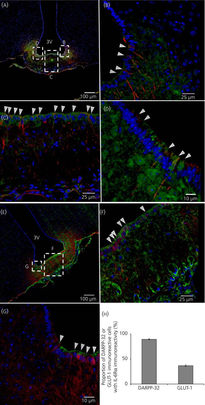
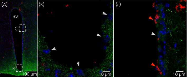
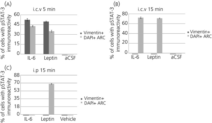
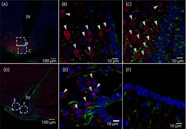
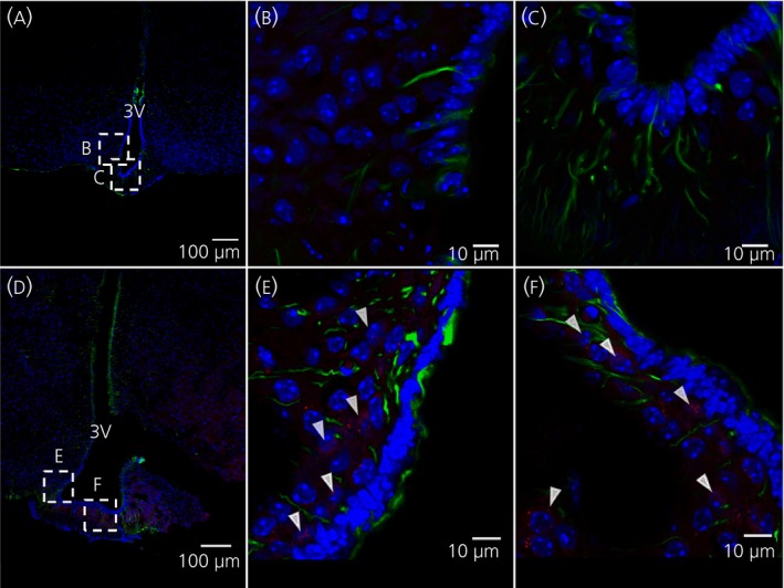
Interleukin (IL)-6- /- mice develop mature onset obesity, whereas i.c.v. injection of IL-6 decreases obesity in rodents. Moreover, levels of IL-6 in cerebrospinal fluid (CSF) were reported to be inversely correlated with obesity in humans. Tanycytes lining the base of the third ventricle (3V) in the hypothalamus have recently been reported to be of importance for metabolism. In the present study, we investigated whether tanycytes could respond to IL-6 in the CSF. With immunohistochemistry using a well characterised antibody directed against the ligand binding receptor for IL-6, IL-6 receptor α (IL-6Rα), it was found that tanycytes, identified by the two markers, vimentin and dopamine- and cAMP-regulated phosphoprotein of 32 kDa, contained IL-6Rα. There were fewer IL-6Rα on another type of ventricle-lining cells, ependymal cells, as identified by the marker glucose transporter-1. To demonstrate that the immunoreactive IL-6Rα were responsive to IL-6, we injected IL-6 i.c.v. This treatment increased immunoreactive phosphorylated signal transducer and activator of transcription-3 (pSTAT3) in tanycytes after 5 minutes and in cells in the medial part of the arcuate nucleus after 5 and 15 minutes. Intracerebroventricular injection of leptin exerted similar effects. As expected, i.p. injection of leptin also induced pSTAT3 staining in the hypothalamus, whereas i.p. IL-6 injection had little effect on this parameter. Intracerebroventricular or i.p. injection of vehicle only had no effect on pSTAT3-immunoreactivity. In summary, there are functional IL-6Rα on tanycytes at the bottom of the 3V, in agreement with the possibility that ventricular administration of IL-6 decreases obesity in mice via an effect on this cell type.
Keywords: IL-6; IL-6Rα; hypothalamus; tanycytes; third ventricle.
© 2017 The Authors. Journal of Neuroendocrinology published by John Wiley & Sons Ltd. on behalf of British Society for Neuroendocrinology.
Figures

References
-
- Hodge D, Hurt E, Farrar W. The role of IL‐6 and STAT3 in inflammation and cancer. Eur J Cancer. 2005;41:2502‐2512. - PubMed
-
- Ishihara K, Hirano T. IL‐6 in autoimmune disease and chronic inflammatory proliferative disease. Cytokine Growth Factor Rev. 2002;13:357‐368. - PubMed
-
- Mauer J, Denson J, Brüning J. Versatile functions for IL‐6 in metabolism and cancer. Trends Immunol. 2015;36:92‐101. - PubMed
-
- Pedersen B, Febbraio M. Muscle as an endocrine organ: focus on muscle‐derived interleukin‐6. Physiol Rev. 2008;88:1379‐1406. - PubMed
-
- Kamimura D, Ishihara K, Hirano T. IL‐6 signal transduction and its physiological roles: the signal orchestration model. Rev Physiol Biochem Pharmacol. 2003;149:1‐38. - PubMed
Publication types
MeSH terms
Substances
LinkOut - more resources
Full Text Sources
Other Literature Sources
Research Materials
