Multicenter Study
. 2018 Jan;39(1):171-188.
doi: 10.1002/hbm.23834. Epub 2017 Oct 11.

Interbrain phase synchronization during turn-taking verbal interaction-a hyperscanning study using simultaneous EEG/MEG

Affiliations
Multicenter Study

Interbrain phase synchronization during turn-taking verbal interaction-a hyperscanning study using simultaneous EEG/MEG

Sangtae Ahn et al. Hum Brain Mapp. 2018 Jan.

Abstract

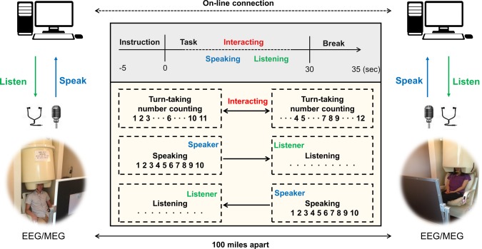
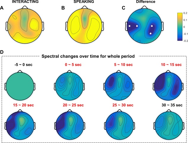
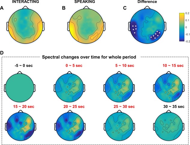
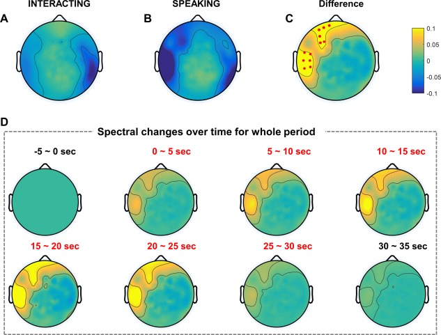
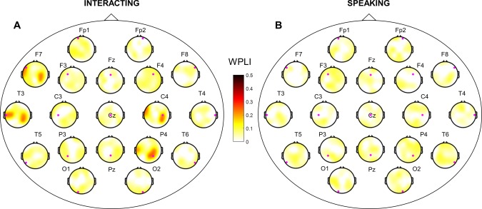
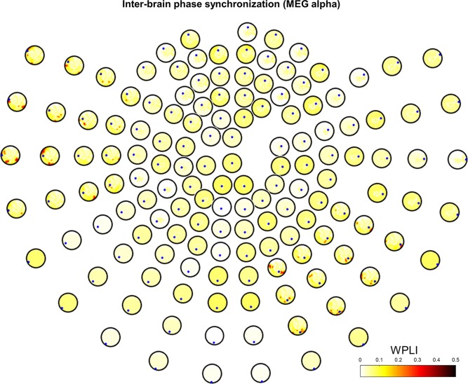
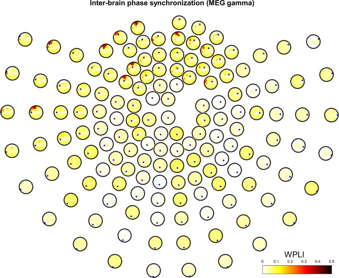
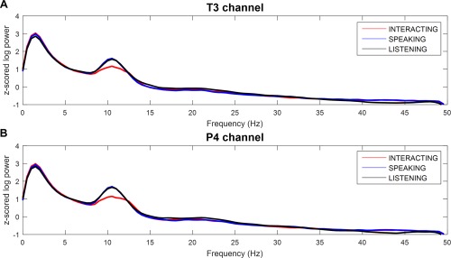
Recently, neurophysiological findings about social interaction have been investigated widely, and hardware has been developed that can measure multiple subjects' brain activities simultaneously. These hyperscanning studies have enabled us to discover new and important evidences of interbrain interactions. Yet, very little is known about verbal interaction without any visual input. Therefore, we conducted a new hyperscanning study based on verbal, interbrain turn-taking interaction using simultaneous EEG/MEG, which measures rapidly changing brain activities. To establish turn-taking verbal interactions between a pair of subjects, we set up two EEG/MEG systems (19 and 146 channels of EEG and MEG, respectively) located ∼100 miles apart. Subjects engaged in verbal communication via condenser microphones and magnetic-compatible earphones, and a network time protocol synchronized the two systems. Ten subjects participated in this experiment and performed verbal interaction and noninteraction tasks separately. We found significant oscillations in EEG alpha and MEG alpha/gamma bands in several brain regions for all subjects. Furthermore, we estimated phase synchronization between two brains using the weighted phase lag index and found statistically significant synchronization in EEG and MEG data. Our novel paradigm and neurophysiological findings may foster a basic understanding of the functional mechanisms involved in human social interactions. Hum Brain Mapp 39:171-188, 2018. © 2017 Wiley Periodicals, Inc.

Keywords: hyperscanning; phase synchronization; simultaneous EEG/MEG; social interaction; turn-taking verbal interaction.


Figures

Figure 1
Figure 1
Experimental setup in turn‐taking verbal interaction using simultaneous EEG/MEG. Verbal communication between subjects was conducted using a condenser microphone and earphones in an online environment. There are three types of tasks: interacting, speaking, and listening. Interacting: counting numbers in turn from 1 to time limit. Speaking: each subject speaks the numbers alone from 1 to time limit without any listening and visual input. Listening: each subject listens to his/her partner's number counting from 1 to time limit without any response. The distance between the two rooms was ∼100 miles. [Color figure can be viewed at http://wileyonlinelibrary.com]
Figure 2
Figure 2
EEG grand‐averaged topography in the alpha band of two tasks. Each topography indicates the normalized log power difference in the interacting (A) and speaking (B) tasks compared to baseline. (C) Topography map shows the difference between the two tasks (interacting, speaking). Small white dots represent electrodes that exhibited statistically significant differences (P < 0.05) with FDR correction. (D) EEG grand‐averaged topography of normalized power differences in the alpha band (interacting–speaking) over time. Baseline: −5–0 s; task: 0–30 s; break: 30–35 s. [Color figure can be viewed at http://wileyonlinelibrary.com]
Figure 3
Figure 3
MEG grand‐averaged topography in the alpha band of two tasks. Each topography indicates the normalized log power difference in the interacting (A) and speaking (B) tasks compared to baseline. (C) The topography map shows the difference between the two tasks (interacting, speaking). Small white dots represent sensors that exhibited statistically significant differences (P < 0.05) with FDR correction. (D) MEG grand‐averaged topography of normalized power differences in the alpha band (interacting, speaking) over time. Baseline: −5–0 s; task: 0–30 s; break: 30–35 s. [Color figure can be viewed at http://wileyonlinelibrary.com]
Figure 4
Figure 4
MEG grand‐averaged topography in the gamma band of two tasks. Each topography indicates the normalized log power differences in the interacting (A) and speaking (B) tasks compared to baseline. (C) The topography map shows the difference between the two tasks (interacting, speaking). Small red dots represent the sensors that exhibited statistically significant differences (P < 0.05) with FDR correction. (D) MEG grand‐averaged topography of normalized power differences in the gamma band (interacting, speaking) over time. Baseline: −5–0 s; task: 0–30 s; break: 30–35 s. [Color figure can be viewed at http://wileyonlinelibrary.com]
Figure 5
Figure 5
Interbrain phase synchronization in EEG alpha band. WPLI for all pairs of synchronization in the two different tasks (interacting, speaking). WPLI across subjects was represented based on statistical testing (1,000 surrogate data, P < 0.05 with FDR correction). (A) and (B) are topographies of interbrain phase synchronization in the interacting and speaking tasks, respectively. Small magenta dots represent seeds from the initiator. [Color figure can be viewed at http://wileyonlinelibrary.com]
Figure 6
Figure 6
Interbrain phase synchronization in MEG alpha band. Grand‐averaged WPLI is represented based on statistical testing (1,000 surrogate data, P < 0.05 with FDR correction) in the interacting task. Small blue dots represent seeds from the initiator. [Color figure can be viewed at http://wileyonlinelibrary.com]
Figure 7
Figure 7
Interbrain phase synchronization in MEG gamma band. Grand‐averaged WPLI is represented based on statistical testing (1,000 surrogate data, P < 0.05 with FDR correction) in the interacting task. Small blue dots represent seeds from the initiator. [Color figure can be viewed at http://wileyonlinelibrary.com]
Figure 8
Figure 8
Comparison of power spectral density for each conditions. (A) Left temporal (T3 channel) and (B) right centro‐parietal (P4 channel) regions in the three different tasks (interacting, speaking, and listening). Power suppression around the alpha band (8–12 Hz) was obtained in the interacting compared to the speaking and listening tasks. [Color figure can be viewed at http://wileyonlinelibrary.com]

References

    1. Ahn M, Ahn S, Hong JH, Cho H, Kim K, Kim BS, Chang JW, Jun SC (2013): Gamma band activity associated with BCI performance: Simultaneous MEG/EEG study. Front Hum Neurosci 7:848. - PMC - PubMed
    1. Anders S, Heinzle J, Weiskopf N, Ethofer T, Haynes J‐D (2011): Flow of affective information between communicating brains. NeuroImage 54:439–446. - PMC - PubMed
    1. Astolfi L, Toppi J, Fallani FDV, Vecchiato G, Salinari S, Mattia D, Cincotti F, Babiloni F (2010): Neuroelectrical hyperscanning measures simultaneous brain activity in humans. Brain Topogr 23:243–256. - PubMed
    1. Astolfi L, Toppi J, D, Vico Fallani F, Vecchiato G, Cincotti F, Wilke CT, Yuan H, Mattia D, Salinari S, He B, Babiloni F (2011): Imaging the social brain by simultaneous hyperscanning during subject interaction. IEEE Intell Syst 26:38–45. - PMC - PubMed
    1. Babiloni F, Astolfi L (2014): Social neuroscience and hyperscanning techniques: Past, present and future. Neurosci Biobehav Rev 44:76–93. - PMC - PubMed

Publication types

LinkOut - more resources