. 2018 Mar;12(3):e1623-e1635.
doi: 10.1002/term.2589. Epub 2017 Nov 28.

Bioengineered lungs generated from human iPSCs-derived epithelial cells on native extracellular matrix

Affiliations

Bioengineered lungs generated from human iPSCs-derived epithelial cells on native extracellular matrix

Mahboobe Ghaedi et al. J Tissue Eng Regen Med. 2018 Mar.

Abstract

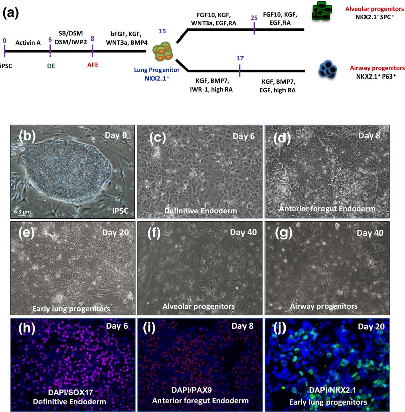
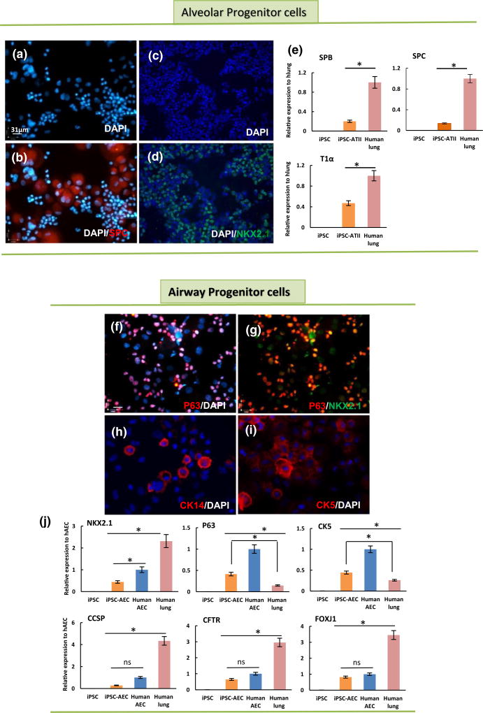
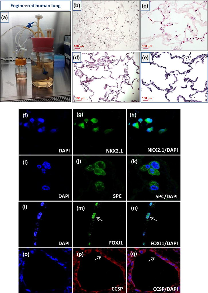
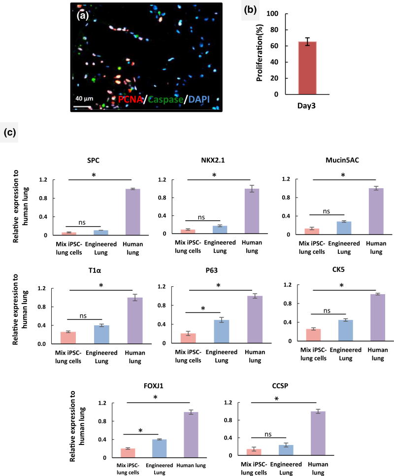
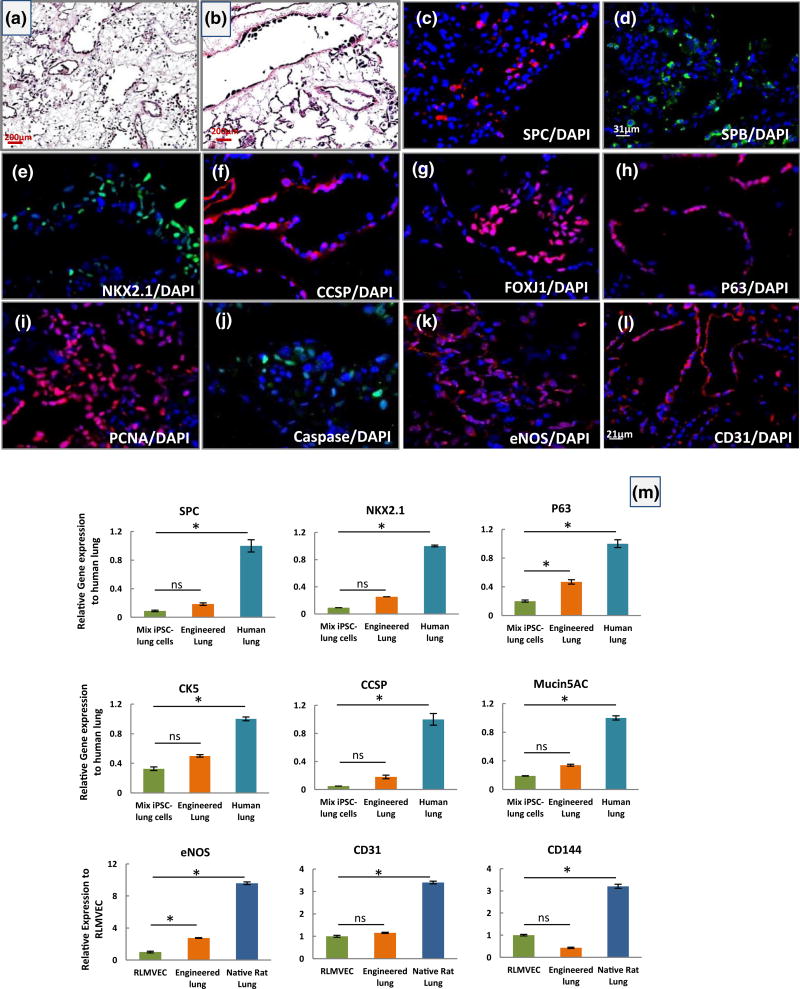
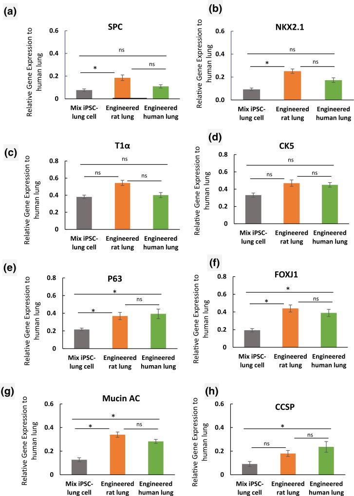
The development of an alternative source for donor lungs would change the paradigm of lung transplantation. Recent studies have demonstrated the potential feasibility of using decellularized lungs as scaffolds for lung tissue regeneration and subsequent implantation. However, finding a reliable cell source and the ability to scale up for recellularization of the lung scaffold still remain significant challenges. To explore the possibility of regeneration of human lung tissue from stem cells in vitro, populations of lung progenitor cells were generated from human iPSCs. To explore the feasibility of producing engineered lungs from stem cells, we repopulated decellularized human lung and rat lungs with iPSC-derived epithelial progenitor cells. The iPSCs-derived epithelial progenitor cells lined the decellularized human lung and expressed most of the epithelial markers when were cultured in a lung bioreactor system. In decellularized rat lungs, these human-derived cells attach and proliferate in a manner similar to what was observed in the decellularized human lung. Our results suggest that repopulation of lung matrix with iPSC-derived lung epithelial cells may be a viable strategy for human lung regeneration and represents an important early step toward translation of this technology.

Keywords: decellularized lung; epithelial cells; induced pluripotent stem cell (iPSC); lung implant; tissue regeneration bioreactor.


Figures

FIGURE 1
FIGURE 1
Generation of lung epithelial cells from human induced pluripotent stem cell (iPSC) in vitro. (a) Schematic for differentiation protocol of human iPSC to alveolar and airway progenitor cells in vitro. (b) Phase-contrast images of human iPSC, (c) definitive endoderm (DE), (d) anterior foregut endoderm (AFE), (e) early lung progenitor cells at day 20, (f–g) alveolar and airway progenitor cells at day 40 (scale bar = 6.3 µm applies to panels b–g). (h–j) immunofluorescent images of differentiated cells from iPSC for (h) SOX17 (endoderm marker) at day 6, (i) PAX9 (anterior foregut endoderm marker) at day 8 and NKX2.1, early marker of lung progenitor cells at day 20 (scale bar = 31 µm applies to panels h–i and 21 µm to panel j). DAPI = 4′,6-diamidino-2-phenylindole; EGF = epidermal growth factor; KGF = keratinocyte growth factor; FGF10 = fibroblast growth factor 10
FIGURE 2
FIGURE 2
Generation of lung alveolar and airway epithelial progenitor cells from human induced pluripotent stem cell (iPSC) in vitro. (a-d) immunofluorescent images of iPSC-alveolar progenitor cells at day 40 (cytocentrifuged prepared), illustrating positive staining for (a) 4′,6-diamidino-2-phenylindole (DAPI), (b) DAPI-SPC, (c) DAPI, and (d) DAPI-NKX2.1. (e) Quantitative reverse transcription-PCR (qRT-PCR) analysis of type II epithelial cell markers in iPSC-lung progenitor cells at day 40; surfactant proteins B and C, and T1α (a type I cell marker). (f–i) immunofluorescent staining for airway markers in iPSC-airway progenitor cells (cytocentrifuged prepared) at day 40: (f) DAPI-P63, (g) NKX2.1 and P63, (h) DAPI-CK14, (i) DAPI-CK5 (scale bar = 31 µm applies to panels a–d and f–i). (j) qRT-PCR analysis of airway epithelial cell markers in iPSC-lung progenitor cells at day 40. For qRT-PCR, values from three independent experiments from the triplicate PCR reaction for each gene were normalized to average glyceraldehyde-3-phosphate dehydrogenase Ct values from the same cDNA samples. The relative expression of each gene was then calculated to normal human lung (hlung) and airway cells isolated from human trachea (hAEC; n = 3 independent replicates for qRT-PCR, error bars indicate ± SEM and *indicates p < .05 and ns indicates no significant difference). CCSP = Clara cell secretory protein; CFTR = cystic fibrosis transmembrane conductance regulator; hAEC = human airway epithelial cells
FIGURE 3
FIGURE 3
Functional culture of engineered human lung made of human lung scaffold and iPSC-derived lung epithelial progenitor cells in a bioreactor system. (a) Recellularized human lung scaffold with iPSC-lung epithelial progenitor cells in a bioreactor culture system. (b–c) Representative images of haematoxylin and eosin sections of recellularized human lung after 4 days of culture. (d–e) Haematoxylin and eosin images of a native human lung (scale bar = 100 µm applies to panels b–e). (f–q) Immunofluorescent images of recellularized lung tissue at day 4 for (f–h) 4′,6-diamidino-2-phenylindole (DAPI) and NKX2.1, (i–k) DAPI and surfactant protein C (SPC), (l–n) DAPI and Forkhead box protein J1 (FOXJ1), and (o–q) DAPI and Clara cell secretory protein (CCSP; scale bar = 63 µm applies to all panels f–q; arrows show the cells landed in airway express FOXJ1 and CCSP)
FIGURE 4
FIGURE 4
Proliferation and gene expression of induced pluripotent stem cell (iPSC)-derived progenitor cells in repopulated human lung matrix. (a) Immunostaining for proliferating cell nuclear antigen (PCNA; red), caspase (green), and 4′,6-diamidino-2-phenylindole (DAPI) in an engineered human lung at day 4 (scale bar = 40 µm) and (b) quantification of percentage of PCNA+ nuclei (y axis, percentage proliferation based on the number of positive nuclei stained for PCNA compared with total cell numbers in three high power fields). (c) Quantitative reverse transcription-PCR analysis of epithelial markers in recellularized lung tissue with iPSC-lung progenitor cells following 4 days of culture. The expression of lung alveolar and airway epithelial cell markers are normalized to glyceraldehyde-3-phosphate dehydrogenase levels, and relative expression of each gene was calculated to normal human lung tissue. (n = 3 tissue samples analysed/lung for quantitative reverse transcription-PCR and error bars indicate ± SEM, *indicates p < .05 and ns indicates no significant difference). CCSP = Clara cell secretory protein; FOXJ1 = Forkhead box protein J1; PCNA = proliferating cell nuclear antigen; SPC = surfactant protein C
FIGURE 5
FIGURE 5
Characterization of engineered rat lung from human iPSC-lung epithelial progenitor cells cultured in the rat bioreactor system. (a–b) Representative H&E staining of engineered rat lung after 4 days of culture (scale bar = 200 µm for a–b panels). (c–l) Immunofluorescence analysis of human lung epithelial cell markers and rat endothelial cell markers in an engineered rat lung at day 4 of culture. (c) surfactant protein C (SPC), (d) surfactant protein B (SPB), (e) NKX2.1, (f) Clara cell secretory protein (CCSP), (g) Forkhead box protein J1 (FOXJ1), (h) P63, (i) proliferating cell nuclear antigen (PCNA), (j) caspase (scale bar = 31 µm applies to all panels except panel l), (k) endothelial nitric oxide synthase (eNOS), and (l) CD31 (scale bar = 21 µm). (m) Quantitative reverse transcription-PCR analysis of human lung alveolar (NKX2.1 and SPC) and airway (P63, CK5, CCSP, and Mucin 5 AC) epithelial markers and of rat endothelial cell markers (eNOS, CD31, and CD144) at day 4 of culture. The Ct values of each gene were normalized to glyceraldehyde-3-phosphate dehydrogenase, and the relative expression of each gene was calculated to the human or rat lung tissue control. (All error bars represent ± SEM. *indicates p < .05 and ns indicates no significant difference)
FIGURE 6
FIGURE 6
Assessment of expression of lung epithelial markers in rat and human lung scaffolds. (a–c) Quantitative reverse transcription-PCR analysis of human lung alveolar epithelial markers (a) surfactant protein C (SPC), (b) NKX2.1, (c) T1α, and (d–h) human lung airway epithelial markers (d) CK5, (e)P63, (f) Forkhead box protein J1 (FOXJ1), (g) Mucin 5 AC, and (h) Clara cell secretory protein (CCSP) in iPSC-derived lung progenitor cells, cultured into the decellularized rat and human lung scaffold compared with native human lung. The expression of each lung epithelial marker was normalized to glyceraldehyde-3-phosphate dehydrogenase levels and relative gene expression to normal human lung tissue were calculated for each gene. (n = 3 of iPSC-lung cell samples before seeding, engineered rat and human lung samples were analysed. Error bars represent the ±SEM, *indicates p < .05 and ns indicates no significant difference)

References

    1. Ali NN, Edgar AJ, Samadikuchaksaraei A, Timson CM, Romanska HM, Polak JM, Bishop AE. Derivation of type II alveolar epithelial cells from murine embryonic stem cells. Tissue Engineering. 2002;8(4):541–550. - PubMed
    1. Badylak SF, Weiss DJ, Caplan A, Macchiarini P. Engineered whole organs and complex tissues. Lancet. 2012;379(9819):943–952. - PubMed
    1. Balestrini JL, Gard AL, Liu A, Leiby KL, Schwan J, Kunkemoeller B, Niklason LE. Production of decellularized porcine lung scaffolds for use in tissue engineering. Integrative Biology (Camb) 2015;7(12):1598–1610. https://doi.org/10.1039/c5ib00063g. - DOI - PMC - PubMed
    1. Beers MF, Moodley Y group RSCW. When is an alveolar type 2 cell an alveolar type 2 cell? A conundrum for lung stem cell biology and regenerative medicine. American Journal of Respiratory Cell and Molecular Biology. 2017 https://doi.org/10.1165/rcmb.2016-0426PS. [Epub ahead of print] - DOI - PMC - PubMed
    1. Booth AJ, Hadley R, Cornett AM, Dreffs AA, Matthes SA, Tsui JL, White ES. Acellular normal and fibrotic human lung matrices as a culture system for in vitro investigation. American Journal of Respiratory and Critical Care Medicine. 2012;186(9):866–876. - PMC - PubMed

Publication types