. 2017 Dec;6(12):2135-2145.
doi: 10.1002/sctm.17-0130. Epub 2017 Oct 10.

Placenta-Derived Adherent Stromal Cells Improve Diabetes Mellitus-Associated Left Ventricular Diastolic Performance

Affiliations

Placenta-Derived Adherent Stromal Cells Improve Diabetes Mellitus-Associated Left Ventricular Diastolic Performance

Sophie Van Linthout et al. Stem Cells Transl Med. 2017 Dec.

Abstract

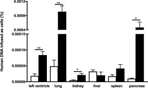
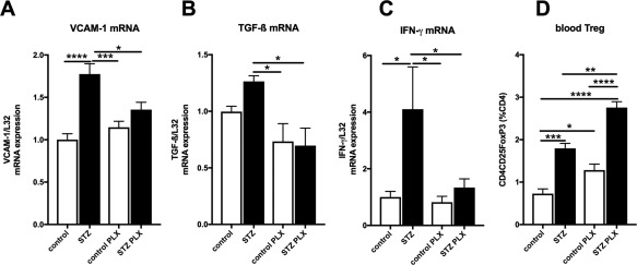
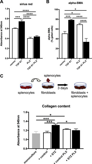
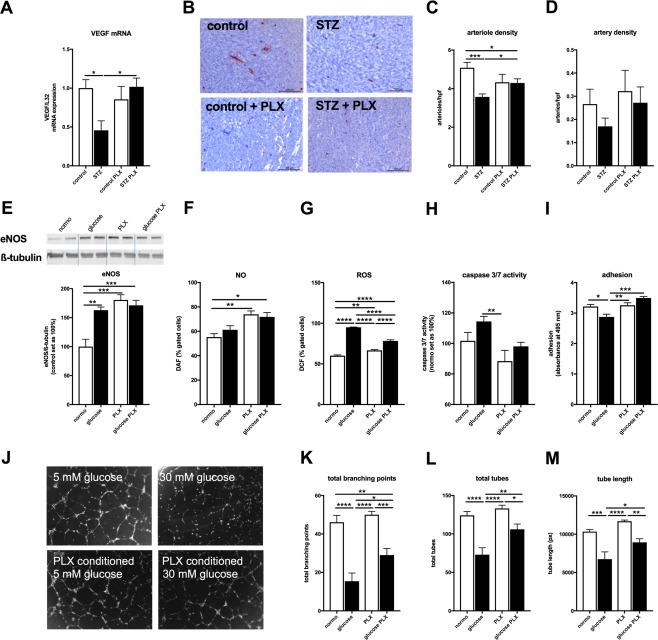
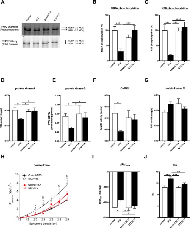
Left ventricular (LV) diastolic dysfunction is among others attributed to cardiomyocyte stiffness. Mesenchymal stromal cells (MSC) have cardiac-protective properties. We explored whether intravenous (i.v.) application of PLacenta-eXpanded (PLX) MSC-like cells (PLX) improves LV diastolic relaxation in streptozotocin (STZ)-induced diabetic mice and investigated underlying mechanisms. Diabetes mellitus was induced by STZ application (50 mg/kg body weight) during five subsequent days. One week after the first STZ injection, PLX or saline were i.v. applied. Two weeks later, mice were hemodynamically characterized and sacrificed. At this early stage of diabetic cardiomyopathy with low-grade inflammation and no cardiac fibrosis, PLX reduced LV vascular cell adhesion molecule-1, transforming growth factor-β1, and interferon-γ mRNA expression, induced the percentage of circulating regulatory T cells, and decreased the splenic pro-fibrotic potential in STZ mice. STZ + PLX mice exhibited higher LV vascular endothelial growth factor mRNA expression and arteriole density versus STZ mice. In vitro, hyperglycemic PLX conditioned medium restored the hyperglycemia-impaired tube formation and adhesion capacity of human umbelical vein endothelial cells (HUVEC) via increasing nitric oxide (NO) bioavailability. PLX further induced the diabetes-downregulated activity of the NO downstream protein kinase G, as well as of protein kinase A, in STZ mice, which was associated with a raise in phosphorylation of the titin isoforms N2BA and N2B. Concomitantly, the passive force was lower in single isolated cardiomyocytes from STZ + PLX versus from STZ mice, which led to an improvement of LV diastolic relaxation. We conclude that i.v. PLX injection improves diabetes mellitus-associated diastolic performance via decreasing cardiomyocyte stiffness. Stem Cells Translational Medicine 2017;6:2135-2145.

Keywords: Diabetes mellitus; Diastole; Passive force; Placenta-expanded cell; Titin.


Figures

Figure 1
Figure 1
Engraftment of PLacenta‐eXpanded (PLX) 14 days after intravenous injection. Bar graphs represent human DNA infused as cells (%) in the left ventricle, lung, kidney, liver, spleen, and pancreas of control mice (open bars) and streptozotocin (STZ)‐treated mice (closed bars) i.v. injected with PLX. For comparison of PLX presence per respective tissue of control versus STZ mice, an unpaired Student's t test was used with *, p < .05; **, p < .005; n = 4/group.
Figure 2
Figure 2
PLX reduce cardiac inflammation and increase blood regulatory T cells in streptozotocin‐induced diabetic mice. Bar graphs represent the mean ± SEM of LV. (A): VCAM‐1, (B): TGF‐β, (C): IFN‐γ, with n = 7–8/PBS groups and n = 5–6/PLX groups, and (D): blood Treg cells, depicted as CD4CD25FoxP3 (% of CD4 cells) with n = 4/group in control (open bars) and STZ (closed bars) mice injected with PBS or PLX, as indicated. For all data: *, p < .05; **, p < .01; ***, p < .001; ****, p < .0001. Abbreviations: IFN‐γ, interferon‐γ; PLX, placenta‐expanded cell; STZ, streptozotocin; TGF‐β1, transforming growth factor‐β1; Treg, regulatory T cells; VCAM, vascular cell adhesion molecule.
Figure 3
Figure 3
PLX exert antifibrotic effects in vitro. PLX conditioned medium reduces TGF‐β1‐induced collagen deposition and α‐SMA expression in cardiac fibroblasts. Bar graphs represent the mean ± SEM of (A) the absorbance at 540 nm detected after Sirus Red staining, depicting collagen deposition, and (B) α‐SMA expression of cardiac fibroblasts cultured in PLX medium, PLX medium + TGF‐β1, PLX conditioned (cond.) medium, or PLX cond. medium + TGF‐β1 as indicated, with open bars representing without TGF‐β1 and black bars with TGF‐β1 conditions, and n = 6/group. Splenocytes derived from STZ PLX mice induce lower collagen deposition in murine fibroblasts upon coculture compared with splenocytes from STZ mice. (C): Schematic representation of the experimental set‐up. (D): Bar graph represents the mean ± SEM of the absorbance at 540 nm detected after Sirus Red staining of fibroblasts cultured in monoculture or cocultured with splenocytes from control, STZ, control PLX, or STZ PLX mice as indicated, with n = 5–6/group. For all data: *, p < .05; **, p < .01; ***, p < .001; ****, p < .0001. Abbrevaitions: PLX, PLacenta‐eXpanded cell; STZ, streptozotocin; TGF‐ß1, transforming growth factor‐β1.
Figure 4
Figure 4
PLX improve hyperglycemia‐impaired vascularization and endothelial function. (A): Bar graphs represent the mean ± SEM of LV VEGF mRNA expression in control (open bars) and STZ (closed bars) mice injected with PBS or PLX, as indicated with n = 7/control and n = 6/STZ and PLX groups. (B): Representative pictures at magnification ×100 of arteries and arterioles in the LV of control, STZ, control + PLX, and STZ + PLX mice as indicated. Bar graphs represent the mean ± SEM of LV (C) arteriole and (D) artery density per high power field (hpf) in control (open bars) and STZ (closed bars) mice injected with PBS or PLX, as indicated with n = 8/control, n = 10/STZ, n = 5/control + PLX, and n = 11/STZ + PLX groups. In vitro, PLX‐conditioned hyperglycemic medium modulate (E) eNOS expression, (F) NO, (G) ROS levels, and (H) caspase 3/7 activity in HUVEC, which is translated in improved (I). adhesion capacity and (J–M) tube formation versus HUVEC cultured under hyperglycemic conditions. Bar graphs represent the mean ± SEM of (E) eNOS expression normalized to β‐tubulin and the control groups set as 100%, (F) % of DAF‐positive, (G) % of DCF‐positive gated cells, with all n = 4/condition, (H) caspase 3/7 activity depicted with the normoglycemia group set as 100% with n = 8–10/condition, and (I) adhered crystal violet‐stained HUVEC depicted as the absorbance at 495 nm with n = 5–6/condition. (J): Representative pictures of HUVEC cultured in normo‐ (5 mM glucose), hyperglycemic (30 mM glucose) medium, or normo‐ (5 mM glucose), hyperglycemic (30 mM glucose) conditioned PLX medium, as indicated. Bar graphs represent the mean ± SEM of (K) total branching points, (L) total tubes, and (M) tube length analyzed from n = 7 to 8 fields/condition. For all data: *, p < .05; **, p < .01; ***, p < .001; ****, p < .0001. Abbrevaitions: eNos, endothelial nitric oxide synthase; NO, nitric oxide; ROS, reactive oxygen species, PLX, placenta‐expanded cell; ROS, reactive oxygen species; STZ, streptozotocin.
Figure 5
Figure 5
PLX restore cardiomyocyte stiffness and improve diastolic relaxation in streptozotocin‐induced diabetic mice. (A): Representative images of titin N2BA and N2B phosphoproteins and total proteins in control, STZ, control PLX, and STZ PLX mice, as indicated. Bar graphs represent the mean ± SEM of (B) N2BA phosphorylation and (C) N2B phosphorylation depicted as % with the control set as 100% of control (open bars) and STZ (closed bars) mice injected with PBS or PLX, as indicated with n = 7/control, n = 10/STZ, n = 5–7/control + PLX and n = 7–8/STZ + PLX groups and *, p < .05; **, p < .01; ***, p < .0005; ****, p < .0001 of (D) PKA (ng/µl) in control (open bars) and STZ (closed bars) mice injected with PBS or PLX, as indicated with n = 3/group and *, p < .05; (E) PKG (pmol/min/µg protein) in control (open bars) and STZ (closed bars) mice injected with PBS or PLX, as indicated with n = 4/PBS group, n = 3/PLX group and *, p < .05, (F) CaMKII activity (CaMKII; mU/ml) in control (open bars) and STZ (closed bars) mice injected with PBS or PLX, as indicated with n = 5/control group, n = 9/STZ group, and n = 4/PLX groups, and *, p < .05, (G) PKC (ng/µl) in control (open bars) and STZ (closed bars) mice injected with PBS or PLX, as indicated with n = 3/group and *, p < .05, (H) Passive tension Fpassive (kN/m2) versus sarcomere length (µm) in control (‐•‐), STZ (‐○‐), control + PLX (‐▪‐), and STZ + PLX (‐◻‐), as indicated, with n = 15–19 cardiomyocytes/group and *, p < .05 versus control and STZ + PLX and §, p < .05 versus control + PLX. Bar graphs represent the mean ± SEM of (I) dP/dtmin and (J) Tau with n = 8 and n = 11 for the control and STZ groups, respectively, and *, p < .05; **, p < .01; ***, p <.0005 analyzed by one‐way analysis of variance (ANOVA) without correction for multiple comparison. Abbreviations: PKA, protein kinase A; PKC, protein kinase C; PKG, protein kinase G; PLX, placenta‐expanded cell; STZ, streptozotocin.

References

    1. Van Linthout S, Spillmann F, Riad A et al. Human apolipoprotein A‐I gene transfer reduces the development of experimental diabetic cardiomyopathy. Circulation 2008;117:1563–1573. - PubMed
    1. Tschope C, Walther T, Königer J et al. Prevention of cardiac fibrosis and left ventricular dysfunction in diabetic cardiomyopathy in rats by transgenic expression of the human tissue kallikrein gene. FASEB J 2004;18:828–835. - PubMed
    1. Chen JX, Zeng H, Reese J et al. Overexpression of angiopoietin‐2 impairs myocardial angiogenesis and exacerbates cardiac fibrosis in the diabetic db/db mouse model. Am J Physiol Heart Circ Physiol 2012;302:H1003–H1012. - PMC - PubMed
    1. Farhangkhoee H, Khan ZA, Kaur H et al. Vascular endothelial dysfunction in diabetic cardiomyopathy: Pathogenesis and potential treatment targets. Pharmacol Ther 2006;111:384–399. - PubMed
    1. Kruger M, Babicz K, von Frieling‐Salewsky M et al. Insulin signaling regulates cardiac titin properties in heart development and diabetic cardiomyopathy. J Mol Cell Cardiol 2010;48:910–916. - PubMed

Publication types