. 2017 Nov;6(11):1972-1986.
doi: 10.1002/sctm.17-0059. Epub 2017 Oct 10.

Enhanced Stem Cell Differentiation and Immunopurification of Genome Engineered Human Retinal Ganglion Cells

Affiliations

Enhanced Stem Cell Differentiation and Immunopurification of Genome Engineered Human Retinal Ganglion Cells

Valentin M Sluch et al. Stem Cells Transl Med. 2017 Nov.

Abstract

Human pluripotent stem cells have the potential to promote biological studies and accelerate drug discovery efforts by making possible direct experimentation on a variety of human cell types of interest. However, stem cell cultures are generally heterogeneous and efficient differentiation and purification protocols are often lacking. Here, we describe the generation of clustered regularly-interspaced short palindromic repeats(CRISPR)-Cas9 engineered reporter knock-in embryonic stem cell lines in which tdTomato and a unique cell-surface protein, THY1.2, are expressed under the control of the retinal ganglion cell (RGC)-enriched gene BRN3B. Using these reporter cell lines, we greatly improved adherent stem cell differentiation to the RGC lineage by optimizing a novel combination of small molecules and established an anti-THY1.2-based protocol that allows for large-scale RGC immunopurification. RNA-sequencing confirmed the similarity of the stem cell-derived RGCs to their endogenous human counterparts. Additionally, we developed an in vitro axonal injury model suitable for studying signaling pathways and mechanisms of human RGC cell death and for high-throughput screening for neuroprotective compounds. Using this system in combination with RNAi-based knockdown, we show that knockdown of dual leucine kinase (DLK) promotes survival of human RGCs, expanding to the human system prior reports that DLK inhibition is neuroprotective for murine RGCs. These improvements will facilitate the development and use of large-scale experimental paradigms that require numbers of pure RGCs that were not previously obtainable. Stem Cells Translational Medicine 2017;6:1972-1986.

Keywords: Biotechnology; Cell differentiation; Clustered regularly interspaced short palindromic repeats; Retinal ganglion cells; Stem cells.


Conflict of interest statement

The authors indicated no potential conflicts of interest.

Figures

Figure 1
Figure 1
Generation of a novel retinal ganglion cell reporter stem cell line. (A): Schematic illustration depicting reporter design. CRISPR‐Cas9 was used to target the stop codon of BRN3B in H7 hESCs. A P2A linked tdTomato was added in tandem with a P2A‐THY1.2 to the BRN3B coding sequence. Following translation, the BRN3B transcription factor protein is localized to the nucleus, tdTomato to the cytoplasm, and THY1.2 to the cell surface. (B): PCR test for zygosity. Primers spanning the integration region were used to amplify genomic DNA for comparison between the parental WT H7 line and the isolated E4‐H7 clone. E4‐H7 DNA produced one band of expected integration size and one wildtype band, indicating heterozygosity of the modified locus. (C): Fluorescence microscopy of day 35 differentiated tdTomato+ cells. Scale bars = 100 µm. Abbreviation: WT, wild type.
Figure 2
Figure 2
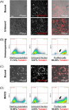
Immunopurification of differentiated stem cell‐derived retinal ganglion cells. (A): Fluorescence and phase microscopy of cells after immunopanning purification. Fluorescent cells bound to anti‐THY1.2 coated plates with high specificity. The unbound fraction contained nonfluorescent cells and cells of lower tdTomato fluorescence intensity. (B): Flow cytometry assessment of the immunopanning method. (C): Fluorescence and phase microscopy of cells after magnetic activated cell sorting (MACS) purification. (D): Flow cytometry assessment of the MACS method. Scale bars = 100 µm. For flow cytometry, red fluorescence intensity is shown on the x‐axis. Differentiated wild type H7 hESCs were used to set a gate threshold for tdTomato fluorescence. Higher fluorescence intensity cells are preferentially retained with the immunopanning method and lower fluorescence intensity cells are lost (black arrow). The MACS method retains more cells of a lower fluorescence intensity compared with immunopanning (black arrow). Abbreviation: BSC‐A, back‐scatter area.
Figure 3
Figure 3
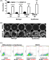
Effects of DSM, Noggin, and IDE2 on retinal ganglion cell differentiation. (A): Flow cytometry analysis of cells treated with DMSO or DSM from day 1 to 8 of differentiation. p value = .0258. (B): Whole‐well fluorescence microscopy of differentiated cells with or without recombinant Noggin treatment for days 1 to 6. (C): Flow cytometry analysis of cells treated with DMSO or IDE2 from day 1 to 6 of differentiation. p value = .3888. (D): Cellomics scan of fluorescent differentiated cultures. IDE2, DSM, or LDN‐193189 alone or in combinations were applied to cultures from day 1 to 4 or 1 to 6. Total fluorescence area was calculated. p values = .9951, .9960, .3145, .0001, and .0001, respectively. All cultures were analyzed on day 40–46. N = 3 where N = independent experiments. *, p < .05; ****, p < .0001. N.S. = not significant. Unpaired two‐tailed t test was used in (A) and (C) and One‐way analysis of variance (ANOVA) (α = 0.05 with Dunnett's multiple comparisons test) was used in (D). Error bars represent standard deviation. Abbreviation: DSM, Dorsomorphin.
Figure 4
Figure 4
Differentiation improvement via addition of DID together with NF and DAPT. (A): Flow cytometry analysis of cells treated with DMSO, DID, NF, or DIDNF on Matrigel coated plates. DID and IDE2 were added from day 1 to 6, NIC from day 1 to 10, FSK from day 1 to 30. p values = .6219, .0431, and .0131, respectively. (B): Flow cytometry analysis of differentiated cells treated with DMSO, DID, NF, or DIDNF on Synthemax coated plates. Whole well fluorescence microscopy images of these cultures are shown in Supporting Information Figure S4d. p values = .9783, .0040, and .0001 for comparisons to DMSO and p value = .0003 for comparison of NF to DIDNF. (C): Flow cytometry analysis of differentiated cells treated with DMSO, DID, or their combination with DAPT added from day 18 to 30. All p values = < .0001. (D): Flow cytometry analysis of differentiated cells treated with DIDNF alone or in combination with DAPT added for the specified time period in days. p values = .0001, .0001, .0001, .0004, .0069, .0009, and .0001, respectively. (E): Schematic of the optimized protocol for RGC differentiation. All cultures were analyzed on day 40–45. N = 3 where N = independent experiments. *, p < .05; **, p < .01; ***, p < .001; ****, p < .0001. N.S. = not significant. ANOVA (α = 0.05) was used in (A–D); Dunnett's multiple comparisons test was used in (A), (B), and (D); Tukey's multiple comparisons test was used in (C) and to compare NF and DIDNF samples in (B). Error bars represent standard deviation. Abbreviations: DAPT, n‐[n‐(3,5‐difluorophenacetyl)‐l‐alanyl]‐s‐phenylglycine t‐butyl ester; DID, Dorsomorphin + IDE2; DIDNF, DID + NF; FSK, forskolin; NIC, nicotinamide; NF, nicotinamide + forskolin.
Figure 5
Figure 5
Validation of small molecule and purification strategy using additional reporter lines. (A): Flow cytometry analysis comparison of DIDNF+D differentiation using the A81‐H7, E4‐H7, and BRN3B‐H9 cell lines on Matrigel and BRN3B‐H9 on Synthemax. BRN3B‐H9 analysis is of cultures shown in (B). p values = <.0001, <.0001, .0008, and .0014, respectively. (B) Whole‐well fluorescence microscopy images of differentiated cells from the BRN3B‐H9 reporter cell line. Cells were treated with either DMSO or DIDNF+D on Matrigel or Synthemax coated plates. (C): Representative flow cytometry analysis of differentiation and subsequent MACS purification of Synthemax cultures in (B). All cultures were analyzed on day 40. For A81‐H7, E4‐H7, and BRN3B‐H9 on Synthemax N = 3. For BRN3B‐H9 on Matrigel N = 4. N = independent experiments. *, p < .05; **, p < .01; ***, p < .001; ****, p < .0001. Unpaired two‐tailed t test was used to compare DMSO versus DIDNF+D for each line in (A). Error bars represent standard deviation. Abbreviations: DIDNF+D = Dorsomorphin + IDE2 + Nicotinamide + Forskolin + DAPT; MACS, magnetic activated cell sorting.
Figure 6
Figure 6
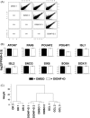
Gene expression analysis of retinal ganglion cell (RGCs) from small molecule driven differentiation. (A): Correlation of gene expression levels between DMSO and DIDNF+D treated samples measured by RNA‐sequencing with two biological replicates per condition. Upper panel: pairwise plotting of log2(FPKM+0.5). FPKM = fragments per kilobase per million mapped reads. Lower panel: Pearson correlation coefficient. (B): Gene expression in DMSO and DIDNF+D treated samples for selected RGC associated genes. (C): Hierarchical clustering of DMSO and DIDNF+D treated samples and GCL and OR collected by laser capture microdissection from human donors. Hierarchical clustering was performed using the Euclidean distance and complete linkage method. Abbreviations: DIDNF+D = Dorsomorphin + IDE2 + Nicotinamide + Forskolin + DAPT; GCL, ganglion cell layer; OR, outer retina.
Figure 7
Figure 7
Stem cell‐derived human retinal ganglion cell (RGCs) have conserved DLK‐dependent injury signaling and are amenable to high‐throughput screening. (A, B): Luminescence‐based survival assay of immunopurified RGCs 48 hours after being challenged with increasing doses of colchicine (A) or a fixed dose of colchicine (1 µM) and increasing doses of tozasertib (B). (C, D): Luminescence‐based survival assay of immunopurified RGCs 48 hours after a colchicine challenge and transfection with increasing doses of control‐ or DLK‐targeting siPOOLs (C) or 1 nM siPOOL (D). Assay validation—Z′ determination. Solid black circles—DLK siPOOL, Open circles—control siPOOL. Shaded areas show three standard deviations from the mean (dashed line) of each population. N =3 where N = independent experiments. Error bars represent standard deviation. Abbreviations: DLK, dual leucine kinase; RLU, relative light units.

References

    1. Kingman S. Glaucoma is second leading cause of blindness globally. Bull World Health Org 2004;82:887–888. - PMC - PubMed
    1. Sluch VM, Zack DJ. Stem cells, retinal ganglion cells and glaucoma. Dev Ophthalmol 2014;53:111–121. - PMC - PubMed
    1. Fernandes KA, Harder JM, Williams PA et al. Using genetic mouse models to gain insight into glaucoma: Past results and future possibilities. Exp Eye Res: 2015;141:42–56. - PMC - PubMed
    1. Levin LA, Gordon LK. Retinal ganglion cell disorders: Types and treatments. Prog Retin Eye Res 2002;21:465–484. - PubMed
    1. Welsbie DS, Yang Z, Ge Y et al. Functional genomic screening identifies dual leucine zipper kinase as a key mediator of retinal ganglion cell death. Proc Natl Acad Sci USA 2013;110:4045–4050. - PMC - PubMed

Publication types

MeSH terms

Substances