Review
. 2017 Oct 11;96(2):267-284.
doi: 10.1016/j.neuron.2017.09.019.

Striatal Local Circuitry: A New Framework for Lateral Inhibition

Affiliations
Review

Striatal Local Circuitry: A New Framework for Lateral Inhibition

Dennis A Burke et al. Neuron. .

Abstract

This Perspective will examine the organization of intrastriatal circuitry, review recent findings in this area, and discuss how the pattern of connectivity between striatal neurons might give rise to the behaviorally observed synergism between the direct/indirect pathway neurons. The emphasis of this Perspective is on the underappreciated role of lateral inhibition between striatal projection cells in controlling neuronal firing and shaping the output of this circuit. We review some classic studies in combination with more recent anatomical and functional findings to lay out a framework for an updated model of the intrastriatal lateral inhibition, where we explore its contribution to the formation of functional units of processing and the integration and filtering of inputs to generate motor patterns and learned behaviors.


Figures

Figure 1
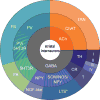
Figure 1. Diversity of cell-types of striatal interneurons
The plot represents the estimated proportions of each subtype of striatal interneuron identified based on their expression of neurotransmitter (inner donut), other molecular markers (middle donut), and electrophysiological properties (outer donut). Abbreviations: ACh, acetylcholine; ChAT, choline-acetyl transferase; TAN, tonically active neurons; TH, tyrosine hydroxlase; CR, calretinin; SOM, somatostatin; NOS, nitric oxide synthase; NPY, neuropeptide Y; LTS, low threshold spiking; NGF, neurogliaform; 5HT3R, serotonin type-3 receptor; FA, fast-adapting; PV, parvalbumin; FS, fast-spiking. *, SOM/NOS LTS neurons are also tonically active, although not classically referred as TANs.
Figure 2
Figure 2. Axo-axonal modulation in the striatum
Simple diagram of known axo-axonal presynaptic modulation by dopamine (DA) and acetylcholine (ACh) in the striatum. D2, dopamine D2 receptor; M5, M5 muscarinic ACh receptor; M2/4, M2 and M4 muscarinic ACh receptors; nAR, nicotinic ACh receptor.
Figure 3
Figure 3. Main identified synaptic connections between local striatal neurons
Schematic showing intrastriatal connectivity between interneurons and projection neurons. Interneurons displayed with molecular markers used to identify cells during experiments. Abbreviations: dSPN, direct pathway projecting medium spiny neuron; iSPN, indirect pathway projecting medium spiny neuron; ChAT, choline-acetyl transferase; TH, tyrosine hydroxlase; SOM, somatostatin; NOS, nitric oxide synthase; NPY, neuropeptide Y; NGF, neurogliaform; 5HT3R, serotonin 3 receptor; PV, parvalbumin;
Figure 4
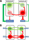
Figure 4. Proposed model of organization of striatal functional units with lateral inhibition within and between units
A, Colored circles represent multiple SPNs from each subclass that are activated together during behavior forming a cluster or ensemble. Each functional unit contains one dSPN cluster and one iSPN cluster. Clusters that are active during execution of behavior A are highlighted and have strong output that enhances behavior A (green arrow) and suppress competing behavior B (red brake). Lateral inhibition from the active clusters further limits activity of the other silent dSPN and iSPN clusters. B, Basic connectivity of the lateral inhibition within and between the two functional units described. Note that all clusters receive excitatory inputs as well. For simplicity, all interneurons are not included in this early conceptual model.
Figure 5
Figure 5. Diverse connectivity patterns for the lateral inhibition generate diverse firing patterns
Left, Diagram of the connectivity pattern for the lateral inhibition within and between the two functional units. Right, raster plots showing the firing of action potentials in response to identical excitatory inputs for each SPN cluster (green for dSPN and red for iSPN clusters) for each functional unit (top raster for FU1, bottom raster for FU2). A, no lateral connections between cells. B, “all-to-all” connections between cells with equal synaptic strengths. C, “all-to-all” connections between cells with asymmetrical connection strengths (iSPN>dSPN). D, proposed organization of lateral inhibition between and within units (“structured”) with equal connection strengths. E, “Structured” connections between cells with asymmetrical connection strengths (iSPN>dSPN).

References

    1. Adler A, Katabi S, Finkes I, Israel Z, Prut Y, Bergman H. Temporal Convergence of Dynamic Cell Assemblies in the Striato-Pallidal Network. 2012;32:2473–2484. doi: 10.1523/JNEUROSCI.4830-11.2012. - DOI - PMC - PubMed
    1. Albin RL, Young AB, Penney JB. The functional anatomy of basal ganglia disorders. Trends Neurosci. 1989;12:366–375. doi: 10.1016/0166-2236(89)90074-X. - DOI - PubMed
    1. Alexander GE, Crutcher MD. Functional architecture of basal ganglia circuits: neural substrates of parallel processing. Trends Neurosci. 1990;13:266–271. doi: 10.1016/0166-2236(90)90107-L. - DOI - PubMed
    1. Alexander GE, DeLong MR, Strick PL. Parallel organization of functionally segregated circuits linking basal ganglia and cortex. Annu. Rev. Neurosci. 1986;9:357–381. doi: 10.1146/annurev.neuro.9.1.357. - DOI - PubMed
    1. Alvarez VA. Clues on the coding of reward cues by the nucleus accumbens. Proc. Natl. Acad. Sci. 2016;113:2560–2562. doi: 10.1073/pnas.1601162113. - DOI - PMC - PubMed

LinkOut - more resources