

Stromal fibroblasts from perimenopausal endometrium exhibit a different transcriptome than those from the premenopausal endometrium
- PMID: 29024986
- PMCID: PMC5803759
- DOI: 10.1093/biolre/iox092
Stromal fibroblasts from perimenopausal endometrium exhibit a different transcriptome than those from the premenopausal endometrium
Abstract
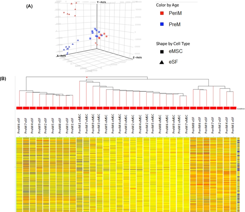
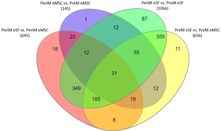
Human endometrium undergoes extensive regeneration on a cyclic basis in premenopausal women and likely occurs through the contribution of stem/progenitor cells. Menopause results in the permanent cessation of menstrual cycles and is preceded by perimenopause, a period of several years in which endocrine and biological changes occur and is a period of risk for endometrial proliferative disorders. The objectives of this study were to identify endometrial mesenchymal stem cells (eMSC) and endometrial stromal fibroblasts (eSF) in endometrium of perimenopausal women and perform expression profile analysis of perimenopausal eMSC and eSF to gain insight into the biology of stem/progenitor and lineage cell populations during the transition to menopause. Endometrial tissue was collected from perimenopausal and premenopausal women (n = 9 each). Microarray analysis was performed on fluorescence-activated cell sorting-isolated eSF and eMSC, and data were validated by quantitative real-time PCR. Principal component analysis showed that cells clustered into three distinct groups in 3-dimensional space: perimenopausal eMSC and premenopausal eMSC clustered together, while perimenopausal eSF and premenopausal eSF formed two discrete clusters separate from eMSC. Hierarchical clustering revealed a branching pattern consistent with principle clustering analysis results, indicating that eMSC from premenopausal and perimenopausal women exhibit similar transcriptomic signatures. Pathway analysis revealed dysregulation of cytoskeleton, proliferation, and survival pathways in perimenopausal vs. premenopausal eSF. These data demonstrate that cell populations have altered gene expression in perimenopausal vs. premenopausal endometrium, and that perimenopausal eSF had altered pathway activation when compared to premenopausal eSF. This study provides insight into aging endometrium with relevance to function in reproductively older women.
Keywords: endometrial mesenchymal stem cell; endometrium; fibroblast; menopause; microarray; small nucleolar RNA.
© The Authors 2017. Published by Oxford University Press on behalf of Society for the Study of Reproduction. All rights reserved. For permissions, please e-mail: journals.permissions@oup.com.
Figures
Similar articles
-
Human Endometrial Fibroblasts Derived from Mesenchymal Progenitors Inherit Progesterone Resistance and Acquire an Inflammatory Phenotype in the Endometrial Niche in Endometriosis.Biol Reprod. 2016 May;94(5):118. doi: 10.1095/biolreprod.115.136010. Epub 2016 Apr 13. Biol Reprod. 2016. PMID: 27075616 Free PMC article.
-
Perivascular human endometrial mesenchymal stem cells express pathways relevant to self-renewal, lineage specification, and functional phenotype.Biol Reprod. 2012 Feb 29;86(2):58. doi: 10.1095/biolreprod.111.095885. Print 2012 Feb. Biol Reprod. 2012. PMID: 22075475 Free PMC article.
-
In vitro evidence that platelet-rich plasma stimulates cellular processes involved in endometrial regeneration.J Assist Reprod Genet. 2018 May;35(5):757-770. doi: 10.1007/s10815-018-1130-8. Epub 2018 Feb 5. J Assist Reprod Genet. 2018. PMID: 29404863 Free PMC article.
-
Endometrial stem/progenitor cells: the first 10 years.Hum Reprod Update. 2016 Mar-Apr;22(2):137-63. doi: 10.1093/humupd/dmv051. Epub 2015 Nov 9. Hum Reprod Update. 2016. PMID: 26552890 Free PMC article. Review.
-
Stem Cells in Endometrial Physiology.Semin Reprod Med. 2015 Sep;33(5):326-32. doi: 10.1055/s-0035-1558405. Epub 2015 Aug 6. Semin Reprod Med. 2015. PMID: 26251119 Review.
Cited by
-
Whole-Tissue Deconvolution and scRNAseq Analysis Identify Altered Endometrial Cellular Compositions and Functionality Associated With Endometriosis.Front Immunol. 2022 Jan 5;12:788315. doi: 10.3389/fimmu.2021.788315. eCollection 2021. Front Immunol. 2022. PMID: 35069565 Free PMC article.
-
Surface Molecular Markers for the Isolation of Viable Fibroblast Subpopulations in the Female Reproductive Tract: A Comprehensive Review.Int J Mol Sci. 2024 Dec 30;26(1):233. doi: 10.3390/ijms26010233. Int J Mol Sci. 2024. PMID: 39796089 Free PMC article. Review.
-
Interleukin-1β induces and accelerates human endometrial stromal cell senescence and impairs decidualization via the c-Jun N-terminal kinase pathway.Cell Death Discov. 2024 Jun 15;10(1):288. doi: 10.1038/s41420-024-02048-6. Cell Death Discov. 2024. PMID: 38879630 Free PMC article.
-
Identification of Potentially Novel Molecular Targets of Endometrial Cancer Using a Non-Biased Proteomic Approach.Cancers (Basel). 2023 Sep 21;15(18):4665. doi: 10.3390/cancers15184665. Cancers (Basel). 2023. PMID: 37760635 Free PMC article.
-
Endometrium as Control of Endometriosis in Experimental Research: Assessment of Sample Suitability.Diagnostics (Basel). 2022 Apr 12;12(4):970. doi: 10.3390/diagnostics12040970. Diagnostics (Basel). 2022. PMID: 35454018 Free PMC article.
References
-
- Gargett CE, Masuda H. Adult stem cells in the endometrium. Mol Hum Reprod 2010; 16:818–834. - PubMed
-
- Du H, Taylor HS. Contribution of bone marrow-derived stem cells to endometrium and endometriosis. Stem Cells 2007; 25:2082–2086. - PubMed
-
- Cervello I, Gil-Sanchis C, Mas A, Simon C. Current understanding of endometrial stem cells. Expert Rev Obstet Gynecol 2009; 4:273–282.
-
- Spitzer TLB, Rojas A, Zelenko Z, Aghajanova L, Erikson DW, Barragan F, Meyer M, Tamaresis JS, Hamilton AE, Irwin JC, Giudice LC. Perivascular human endometrial mesenchymal stem cells express pathways relevant to self-renewal, lineage specification, and functional phenotype. Biol Reprod 2012; 86:58, 1–16. - PMC - PubMed
MeSH terms
Substances
Grants and funding
LinkOut - more resources
Full Text Sources
Other Literature Sources
Molecular Biology Databases
