

Effects of noncavity-distorting fibroids on endometrial gene expression and function
- PMID: 29025102
- PMCID: PMC6279121
- DOI: 10.1093/biolre/iox107
Effects of noncavity-distorting fibroids on endometrial gene expression and function
Abstract
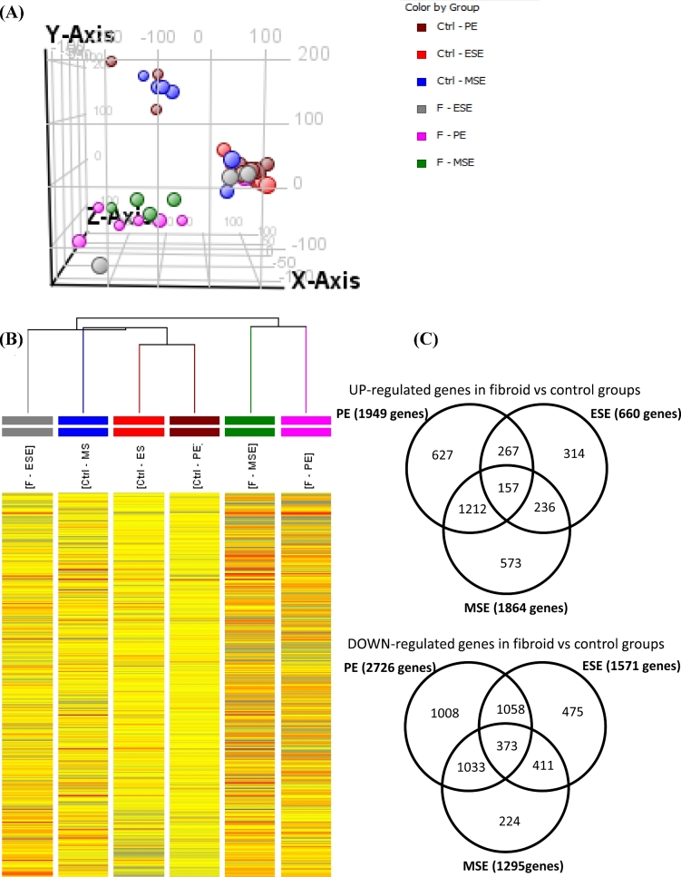
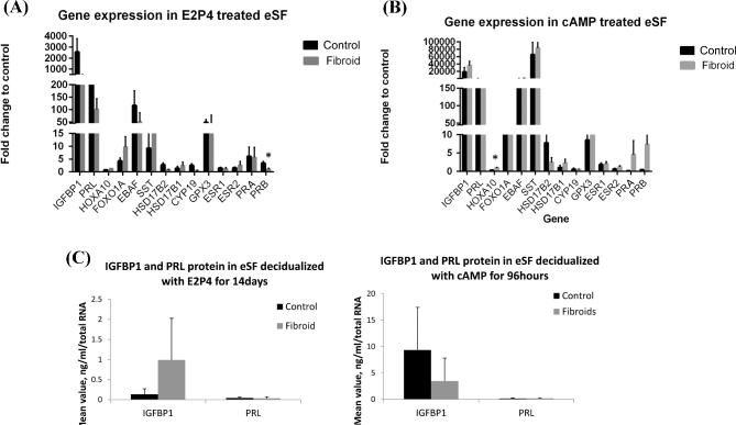
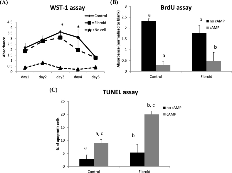
Uterine fibroids are a common finding in infertility patients. Impaired implantation and decidualization have been proposed to contribute to compromised fertility. Data are limited on the endometrial transcriptome from subjects with uterine fibroids, as well as endometrial receptivity and decidualization potential of endometrial stromal fibroblasts (eSF) from women with fibroids. Our objective was to investigate the endometrial transcriptome of women with noncavity-distorting intramural fibroids and compare them to control subjects with no uterine pathology throughout the menstrual cycle. We also evaluated endometrial receptivity gene expression and basic endometrial functions such as decidualization, proliferation, and apoptosis in women with fibroid uterus. Results showed that large numbers of transcripts were significantly dysregulated throughout the menstrual cycle in fibroid subjects compared to controls. However, there were essentially no differences in the expression of receptivity markers at the tissue level, as well as decidualization markers in tissue and eSF in subjects with fibroids compared to controls. However, eSF from women with a fibroid uterus exhibited decreased proliferation potential and increased apoptosis upon decidualization. These data indicate preserved implantation and decidualization potential despite observed gene expression changes in endometrium from women with noncavity-distorting fibroids compared to controls. How this phenomenon and altered proliferation/apoptosis may contribute to impairment of endometrial function in subfertile patients warrants further investigation.
Keywords: apoptosis; decidualization; endometrial fibroblasts; noncavity-distorting fibroids; proliferation; receptivity.
© The Authors 2017. Published by Oxford University Press on behalf of Society for the Study of Reproduction. All rights reserved. For permissions, please e-mail: journals.permissions@oup.com.
Figures
Similar articles
-
Aberrant Akt Activation During Implantation Window in Infertile Women With Intramural Uterine Fibroids.Reprod Sci. 2018 Aug;25(8):1243-1253. doi: 10.1177/1933719117737844. Epub 2017 Nov 7. Reprod Sci. 2018. PMID: 29113583
-
Endometrial Expression of Homeobox Genes and Cell Adhesion Molecules in Infertile Women With Intramural Fibroids During Window of Implantation.Reprod Sci. 2017 Mar;24(3):435-444. doi: 10.1177/1933719116657196. Epub 2016 Jul 19. Reprod Sci. 2017. PMID: 27407137
-
Expression of Endometrial Receptivity Genes Increase After Myomectomy of Intramural Leiomyomas not Distorting the Endometrial Cavity.Reprod Sci. 2016 Jan;23(1):31-41. doi: 10.1177/1933719115612929. Epub 2015 Oct 27. Reprod Sci. 2016. PMID: 26507873
-
Literature Review on the Role of Uterine Fibroids in Endometrial Function.Reprod Sci. 2018 May;25(5):635-643. doi: 10.1177/1933719117725827. Epub 2017 Aug 22. Reprod Sci. 2018. PMID: 28826369 Free PMC article. Review.
-
The Impact of Noncavity-Distorting Intramural Fibroids on Live Birth Rate in In Vitro Fertilization Cycles: A Systematic Review and Meta-Analysis.J Womens Health (Larchmt). 2020 Feb;29(2):210-219. doi: 10.1089/jwh.2019.7813. Epub 2019 Dec 10. J Womens Health (Larchmt). 2020. PMID: 31821069
Cited by
-
A molecular staging model for accurately dating the endometrial biopsy.Nat Commun. 2023 Oct 6;14(1):6222. doi: 10.1038/s41467-023-41979-z. Nat Commun. 2023. PMID: 37798294 Free PMC article.
-
Understanding the Impact of Uterine Fibroids on Human Endometrium Function.Front Cell Dev Biol. 2021 May 25;9:633180. doi: 10.3389/fcell.2021.633180. eCollection 2021. Front Cell Dev Biol. 2021. PMID: 34113609 Free PMC article. Review.
-
Large, Non-Cavity Distorting Intramural Leiomyomas Decrease Leukemia Inhibitory Factor in the Secretory Phase Endometrium.Reprod Sci. 2020 Feb;27(2):569-574. doi: 10.1007/s43032-019-00056-x. Epub 2020 Jan 1. Reprod Sci. 2020. PMID: 32046421 Free PMC article.
-
The role of magnetic resonance-guided focused ultrasound in fertility-sparing treatment of uterine fibroids-current perspectives.Ecancermedicalscience. 2020 May 6;14:1034. doi: 10.3332/ecancer.2020.1034. eCollection 2020. Ecancermedicalscience. 2020. PMID: 32419846 Free PMC article.
-
Uterine Transcriptome: Understanding Physiology and Disease Processes.Biology (Basel). 2023 Apr 21;12(4):634. doi: 10.3390/biology12040634. Biology (Basel). 2023. PMID: 37106834 Free PMC article. Review.
References
-
- Baird DD, Dunson DB, Hill MC, Cousins D, Schectman JM. High cumulative incidence of uterine leiomyoma in black and white women: ultrasound evidence. Am J Obstet Gynecol 2003; 188(1):100–107. - PubMed
-
- Stewart EA. Clinical practice. Uterine fibroids. N Engl J Med 2015; 372(17):1646–1655. - PubMed
-
- Munro MG, Critchley HO, Fraser IS Group FMDW . The FIGO classification of causes of abnormal uterine bleeding in the reproductive years. Fertil Steril 2011; 95(7):2204–2208, 2208 e2201-2203. - PubMed
-
- Olive DL, Pritts EA. Fibroids and reproduction. Semin Reprod Med 2010; 28(3):218–227. - PubMed
-
- Pritts EA, Parker WH, Olive DL. Fibroids and infertility: an updated systematic review of the evidence. Fertil Steril 2009; 91(4):1215–1223. - PubMed
MeSH terms
Grants and funding
LinkOut - more resources
Full Text Sources
Other Literature Sources
