. 2017 Oct 12;6(10):e006126.
doi: 10.1161/JAHA.117.006126.

Compromised Cerebrovascular Regulation and Cerebral Oxygenation in Pulmonary Arterial Hypertension

Affiliations

Compromised Cerebrovascular Regulation and Cerebral Oxygenation in Pulmonary Arterial Hypertension

Simon Malenfant et al. J Am Heart Assoc. .

Abstract

Background: Functional cerebrovascular regulatory mechanisms are important for maintaining constant cerebral blood flow and oxygen supply in heathy individuals and are altered in heart failure. We aim to examine whether pulmonary arterial hypertension (PAH) is associated with abnormal cerebrovascular regulation and lower cerebral oxygenation and their physiological and clinical consequences.

Methods and results: Resting mean flow velocity in the middle cerebral artery mean flow velocity in the middle cerebral artery (MCAvmean); transcranial Doppler), cerebral pressure-flow relationship (assessed at rest and during squat-stand maneuvers; analyzed using transfer function analysis), cerebrovascular reactivity to CO2, and central chemoreflex were assessed in 11 patients with PAH and 11 matched healthy controls. Both groups also completed an incremental ramp exercise protocol until exhaustion, during which MCAvmean, mean arterial pressure, cardiac output (photoplethysmography), end-tidal partial pressure of CO2, and cerebral oxygenation (near-infrared spectroscopy) were measured. Patients were characterized by a significant decrease in resting MCAvmean (P<0.01) and higher transfer function gain at rest and during squat-stand maneuvers (both P<0.05). Cerebrovascular reactivity to CO2 was reduced (P=0.03), whereas central chemoreceptor sensitivity was increased in PAH (P<0.01), the latter correlating with increased resting ventilation (R2=0.47; P<0.05) and the exercise ventilation/CO2 production slope (V˙E/V˙CO2 slope; R2=0.62; P<0.05) during exercise for patients. Exercise-induced increases in MCAvmean were limited in PAH (P<0.05). Reduced MCAvmean contributed to impaired cerebral oxygen delivery and oxygenation (both P<0.05), the latter correlating with exercise capacity in patients with PAH (R2=0.52; P=0.01).

Conclusions: These findings provide comprehensive evidence for physiologically and clinically relevant impairments in cerebral hemodynamic regulation and oxygenation in PAH.

Keywords: central chemoreceptor sensitivity; cerebral ischemia; cerebral oxygenation; cerebrovascular reactivity to CO2; exercise physiology.


Figures

Figure 1
Figure 1
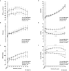
Resting cerebral blood flow (CBF) is reduced in patients with pulmonary arterial hypertension (PAH) compared with controls (Ctrls). Patients with PAH (open circles) exhibited lower resting CBF compared with Ctrls (closed circles), as assessed using systolic (P=0.08) (A), diastolic (P=0.002) (B), and mean flow velocity in the middle cerebral artery (MCAvmean; P=0.004) (C). Consequently, patients with PAH exhibited increased Gosling pulsatility index (D), whereas cerebrovascular conductance index (CVCi; P=0.27) (E) and cerebrovascular resistance index (CVRi; P=0.25) (F) were similar. dMCAv indicates diastolic middle cerebral artery blood flow velocity; and sMCAv, systolic middle cerebral artery blood flow velocity.
Figure 2
Figure 2
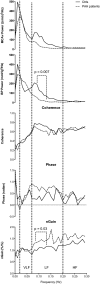
Spontaneous oscillation spectral power and transfer function analysis of resting cerebral blood flow and blood pressure (BP) for the entire spectrum from 0.00 to 0.30 Hz. Continuous (controls [Ctrls]; n=11) and dotted (patients with pulmonary arterial hypertension [PAH]; n=10) lines represent group‐averaged middle cerebral artery blood flow velocity (MCAv) and BP spectral power, coherence, phase, and normalized gain (nGain). HF indicates high frequency (>0.20 Hz); LF, low frequency (0.07–0.20 Hz); and VLF, very low frequency (0.02–0.07 Hz).
Figure 3
Figure 3
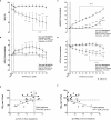
Driven oscillation spectral power and transfer function analysis of cerebral blood flow and blood pressure (BP). Group‐averaged spectral power (A through D), coherence (E), phase (F), and normalized gain (nGain; G) for middle cerebral artery blood flow velocity (MCAv) and BP at 10‐second squat‐stand (0.05 Hz) and at 5‐second squat‐stand (0.10 Hz). Ctrls indicates controls; and PAH, pulmonary arterial hypertension.
Figure 4
Figure 4
Cerebrovascular reactivity to CO 2 is decreased, whereas central chemoreceptor sensitivity is increased in patients with pulmonary arterial hypertension (PAH). Patients with PAH (open circles) exhibited a lower cerebrovascular reactivity to CO 2 (A) and a lower cerebrovascular conductance index (CVCi)–CO 2 reactivity (B) compared with controls (Ctrls; closed circles). No differences were observed between groups on the mean arterial pressure (MAP)–CO 2 reactivity (C). Patients had increased central chemoreceptor sensitivity compared with Ctrls (D). Among patients with PAH, chemoreceptor sensitivity correlated with minute ventilation at rest (E) and with V˙E/V˙CO2 slope during exercise (F). MCAvmean indicates middle cerebral artery mean blood flow velocity; V˙E, minute ventilation; and V˙E/V˙CO2 slope, ventilatory equivalent for CO 2 slope.
Figure 5
Figure 5
Cerebral blood flow and central hemodynamic and ventilatory response in patients with pulmonary arterial hypertension (PAH) and controls (Ctrls) during cardiopulmonary exercise testing. Patients with PAH (open circles) exhibited limited increases in middle cerebral artery mean blood flow velocity (MCAvmean) compared with Ctrls (closed circles) (A, P=0.04), despite similar increases in cardiac output (CO; P=0.96; B). Patients with PAH also had a smaller increase in mean arterial pressure (MAP; P=0.04) compared with Ctrls (C). Although patients with PAH and Ctrls had comparable minute ventilation (VE) at their maximal workload (D), patients with PAH had a higher ventilatory equivalent for CO 2 (V˙E/V˙CO2) (P<0.0001; E) and a persistently lower end‐tidal pressure in CO2 (PETCO 2) compared with Ctrls (P<0.0001; F). The x axis represents the different stages of exercise (rest, unloaded pedaling, and 10%, 20%, 30%, 40%, 50%, 60%, 70%, 80%, 90%, and 100% of maximal workload). The unloaded pedaling is the average of the last 30 seconds before workload onset. Resting MCAvmean, resting hemodynamic and ventilatory responses are calculated as the average of the last stable minute before beginning unloaded pedaling. Each MCAvmean, hemodynamic, and ventilatory response data point represents a 10‐second average for its relative workload. NS indicates nonsignificant; RM, repeated measures. Significantly different from rest: P<0.05, †† P<0.01, †††† P<0.0001. Patients with PAH vs Ctrls: ***P<0.001, ****P<0.0001.
Figure 6
Figure 6
Cerebral oxygen capillary saturation is impaired and correlates with exercise tolerance in pulmonary arterial hypertension (PAH). Patients with PAH (open circles) exhibited a rapid and constant decrease in systemic oxygen saturation (SpO2) at exercise (A), contributing to a significantly lower estimated cerebral oxygen delivery (cDO 2), expressed as changes from baseline (B). Patients with PAH exhibited a sustained decrease in cerebral tissue oxygenation index (∆cTOI), while exhibiting a sustained increased in cerebral deoxyhemoglobin (∆[cHHb]) compared with controls (Ctrls; closed circles; C and D). Lower ∆cTOI and increased ∆[cHHb] correlated with maximal exercise capacity in patients with PAH (E and F). The x axis represents the different stages of exercise (rest, unloaded pedaling, and 10%, 20%, 30%, 40%, 50%, 60%, 70%, 80%, 90%, and 100% of maximal workload). The unloaded pedaling is the average of the last 30 seconds before workload onset. RM indicates repeated measures; and VO2max % pred., predicted value of maximal oxygen consumption. Significantly different from rest: P<0.05, †† P<0.01, ††† P<0.001, †††† P<0.0001. Patients with PAH vs Ctrls: *P<0.05, **P<0.01, ***P<0.001, **** P<0.0001.

References

    1. Galiè N, Humbert M, Vachiery JL, Gibbs S, Lang I, Torbicki A, Simonneau G, Peacock A, Vonk‐Noordegraaf A, Beghetti M, Ghofrani A, Gomez‐Sanchez MA, Hansmann G, Klepetko W, Lancellotti P, Matucci M, McDonagh T, Piérard LA, Trindade PT, Zompatori M, Hoeper M. 2015 ESC/ERS guidelines for the diagnosis and treatment of pulmonary hypertension: the Joint Task Force for the Diagnosis and Treatment of Pulmonary Hypertension of the European Society of Cardiology and the European Respiratory Society. Eur Respir J. 2015;46:903–975. - PubMed
    1. Galiè N, Manes A, Negro L, Palazzini M, Bacchi‐Reggiani ML, Branzi A. A meta‐analysis of randomized controlled trials in pulmonary arterial hypertension. Eur Heart J. 2009;30:394–403. - PMC - PubMed
    1. Lajoie AC, Lauzière G, Lega JC, Lacasse Y, Martin S, Simard S, Bonnet S, Provencher S. Combination therapy versus monotherapy for pulmonary arterial hypertension: a meta‐analysis. Lancet Respir Med. 2016;4:291–305. - PubMed
    1. Rival G, Lacasse Y, Martin S, Bonnet S, Provencher S. Effect of pulmonary arterial hypertension‐specific therapies on health‐related quality of life. Chest. 2014;146:686–708. - PubMed
    1. Benza RL, Miller DP, Barst RJ, Badesch DB, Frost AE, McGoon MD. An evaluation of long‐term survival from time of diagnosis in pulmonary arterial hypertension from reveal. Chest. 2012;142:448–456. - PubMed

MeSH terms