. 2018 Mar;59(3):486-493.
doi: 10.2967/jnumed.117.191221. Epub 2017 Oct 12.

Low-Level Endogenous PSMA Expression in Nonprostatic Tumor Xenografts Is Sufficient for In Vivo Tumor Targeting and Imaging

Affiliations

Low-Level Endogenous PSMA Expression in Nonprostatic Tumor Xenografts Is Sufficient for In Vivo Tumor Targeting and Imaging

Sridhar Nimmagadda et al. J Nucl Med. 2018 Mar.

Abstract

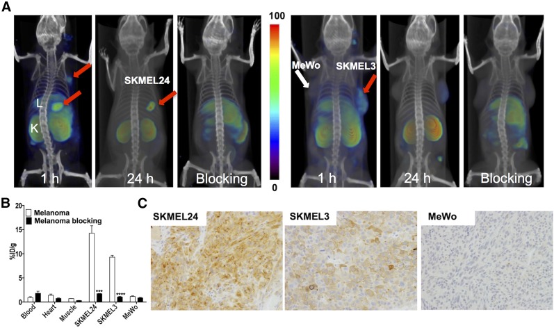
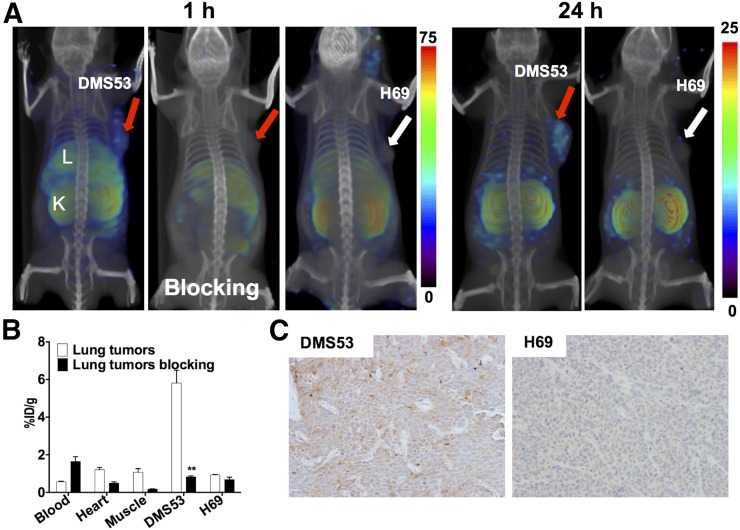
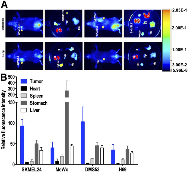
Prostate-specific membrane antigen (PSMA) is highly expressed in prostate cancer and within the neovasculature of other solid tumors. The nonprostatic expression of PSMA has been reported exclusively within the neovasculature endothelial cells of nonprostatic cancers; however, there are few reports on PSMA expression in epithelial cells. Herein, we describe PSMA expression in nonprostatic epithelial cells and characterize the potential of PSMA-binding agents to noninvasively detect that expression. Methods: PSMA expression data were extracted from publicly available genomic databases. Genomic data were experimentally validated for PSMA expression-by quantitative reverse transcription polymerase chain reaction, flow cytometry, and Western blotting-in several nonprostatic cell lines and xenografts of melanoma and small cell lung cancer (SCLC) origin. The feasibility of PSMA detection in those tumor models was further established using PSMA-based nuclear and optical imaging agents and by biodistribution, blocking, and ex vivo molecular characterization studies. Results: We discovered that a small percentage of nonprostatic cancer cell lines and tumors express PSMA. Importantly, PSMA expression was sufficiently high to image established melanoma and SCLC xenografts using PSMA-based nuclear and optical imaging agents. Conclusion: These results indicate that PSMA expression in nonprostatic tumors may not be limited to the endothelium but may also include solid tumor tissue of nonprostatic cancers including melanoma and SCLC. Our observations indicate broader applicability of PSMA-targeted imaging and therapeutics.

Keywords: CCLE; TCGA; lung cancer; melanoma; molecular imaging; prostate cancer.


Figures

FIGURE 1.
FIGURE 1.
PSMA expression in human tumors. (A) FOLH1 gene expression in cell lines extracted from CCLE database, converted into z score, and plotted on the basis of cancer type. Dot plot with line at median value of prostate cancer cell lines is shown. (B) Expression of FOLH1 in different tissue types from TCGA RNA-Seq version 2 data. Dots on top and bottom of box represent outliers. Bars at top and bottom of box represent minimum and maximum expression values of FOLH1 gene, excluding outliers. Box represents 50% of samples. Horizontal bold line inside box is median FOLH1 expression in disease type. CNS = central nervous system; NSCLC = non–small cell lung carcinoma; UADT = upper aerodigestive tract; PRAD = prostate adenocarcinoma; KIRC = kidney renal clear cell carcinoma; LIHC = liver hepatocellular carcinoma; UCEC = uterine corpus endometrial carcinoma; LGG = brain lower-grade glioma; GBM = glioblastoma multiforme; LUSC = lung squamous cell carcinoma; KICH = kidney chromophobe; OV = ovarian carcinoma; BRCA = breast invasive carcinoma; THCA = thyroid carcinoma; HNSC = head and neck squamous cell carcinoma; LUAD = lung adenocarcinoma; SKCM = skin cutaneous melanoma; BLCA = bladder urothelial carcinoma; READ = rectum adenocarcinoma; COAD = colon adenocarcinoma; KIRP = kidney renal papillary cell carcinoma; LAML = acute myeloid leukemia.
FIGURE 2.
FIGURE 2.
Validation of PSMA expression in nonprostatic human cancer cell lines and xenografts. (A) PSMA gene expression in selected melanoma and lung cancer cell lines. (B) Flow cytometry of PSMA surface expression in PSMA-positive PC3 PIP, PSMA-negative PC3 flu, and nonprostatic cancer cell lines. PSMA-positive PC3 PIP is designed to overexpress copious amounts of PSMA. Mean fluorescence intensity quantification shows that PSMA-positive PC3 PIP has high level of PSMA surface expression whereas PSMA-negative PC3 flu, MeWo, and H69 are negative and DMS53, SKMEL24, and SKMEL3 are positive (right panel). (C) PSMA gene expression in selected melanoma and lung cancer xenografts. (D) PSMA total protein levels in selected melanoma and lung cancer xenografts and densitometric quantification (right panel).
FIGURE 3.
FIGURE 3.
PSMA imaging in subcutaneous melanoma xenografts with known PSMA-specific radiotracer, 125I-DCIBzL. (A) Male NOD/SCID mice bearing SKMEL24 xenografts or SKMEL3 and MeWo were injected with 37 MBq (1 mCi) of 125I-DCIBzL through tail vein, and SPECT/CT images were acquired 1 and 24 h later. Arrows = tumor; L = liver; K = kidney. (B) Mice harboring SKMEL24 or SKMEL3 and MeWo xenografts were administered 74 kBq (20 μCi) of 125I-DCIBzL via tail vein injection, and biodistribution studies were performed 1 h afterward. For blocking dose, DCIBzL at 50 mg/kg was injected subcutaneously 30 min before 125I-DCIBzL. Data are mean ± SEM of 4 animals. Significance of value is indicated by asterisks, and comparative reference is blocking dose uptake in same tumor. ***P < 0.001. ****P < 0.0001. (C) Representative microscopic images of PSMA-stained sections from same cohort of mice obtained at ×20 magnification.
FIGURE 4.
FIGURE 4.
PSMA imaging in subcutaneous lung cancer xenografts with known PSMA-specific radiotracer, 125I-DCIBzL. (A) Male NOD/SCID mice bearing DMS53 or H69 xenografts were administered 37 MBq (1 mCi) of 125I-DCIBzL via tail vein injection, and SPECT/CT images were acquired 1 h and 24 h (right panels) afterward. Arrows = tumor; L = liver; K = kidney. (B) Mice harboring DMS53 or H69 xenografts were administered 74 kBq (20 μCi) of 125I-DCIBzL, and biodistribution studies were performed 1 h afterward. For blocking dose, DCIBzL at 50 mg/kg was coinjected with 125I-DCIBzL. Data are mean ± SEM of 4 animals. Significance of value is indicated by asterisks, and comparative reference is blocking dose uptake in same tumor. **P < 0.01. (C) Representative microscopy images of PSMA-stained sections from same cohort of mice obtained at ×20 magnification.
FIGURE 5.
FIGURE 5.
Near-infrared fluorescence imaging of PSMA expression in subcutaneous melanoma and lung xenografts with YC-27. (A) Male NOD/SCID mice bearing melanoma and lung xenografts were administered 1 nmol of a near-infrared–labeled PSMA-targeting reagent, YC-27, via tail vein injection, and fluorescence images were acquired 24 h afterward. (B) After near-infrared imaging, selected tissues were harvested and fluorescence intensity was measured and normalized to muscle fluorescence intensity. Arrow = tumor; H = heart; K = kidney; L = liver; Ms = muscle; Sp = spleen; St = stomach.

References

    1. Davis MI, Bennett MJ, Thomas LM, Bjorkman PJ. Crystal structure of prostate-specific membrane antigen, a tumor marker and peptidase. Proc Natl Acad Sci USA. 2005;102:5981–5986. - PMC - PubMed
    1. Pavlícek J, Ptacek J, Bařinka C. Glutamate carboxypeptidase II: an overview of structural studies and their importance for structure-based drug design and deciphering the reaction mechanism of the enzyme. Curr Med Chem. 2012;19:1300–1309. - PubMed
    1. Bařinka C, Rojas C, Slusher B, Pomper M. Glutamate carboxypeptidase II in diagnosis and treatment of neurologic disorders and prostate cancer. Curr Med Chem. 2012;19:856–870. - PMC - PubMed
    1. Sweat SD, Pacelli A, Murphy GP, Bostwick DG. Prostate-specific membrane antigen expression is greatest in prostate adenocarcinoma and lymph node metastases. Urology. 1998;52:637–640. - PubMed
    1. Bostwick DG, Pacelli A, Blute M, Roche P, Murphy GP. Prostate specific membrane antigen expression in prostatic intraepithelial neoplasia and adenocarcinoma: a study of 184 cases. Cancer. 1998;82:2256–2261. - PubMed

Publication types

Substances