. 2017 Oct 12;7(1):13018.
doi: 10.1038/s41598-017-12530-0.

Crystal structure of the flexible tandem repeat domain of bacterial cellulose synthesis subunit C

Affiliations

Crystal structure of the flexible tandem repeat domain of bacterial cellulose synthesis subunit C

Shingo Nojima et al. Sci Rep. .

Abstract

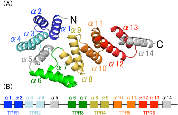
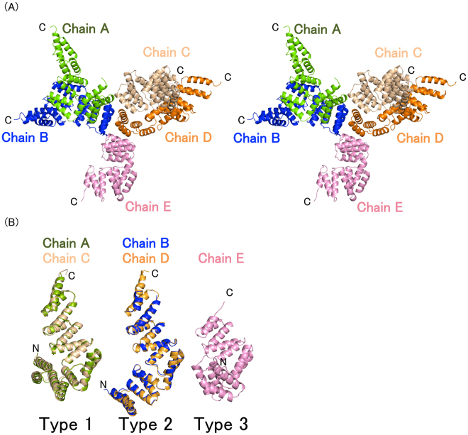
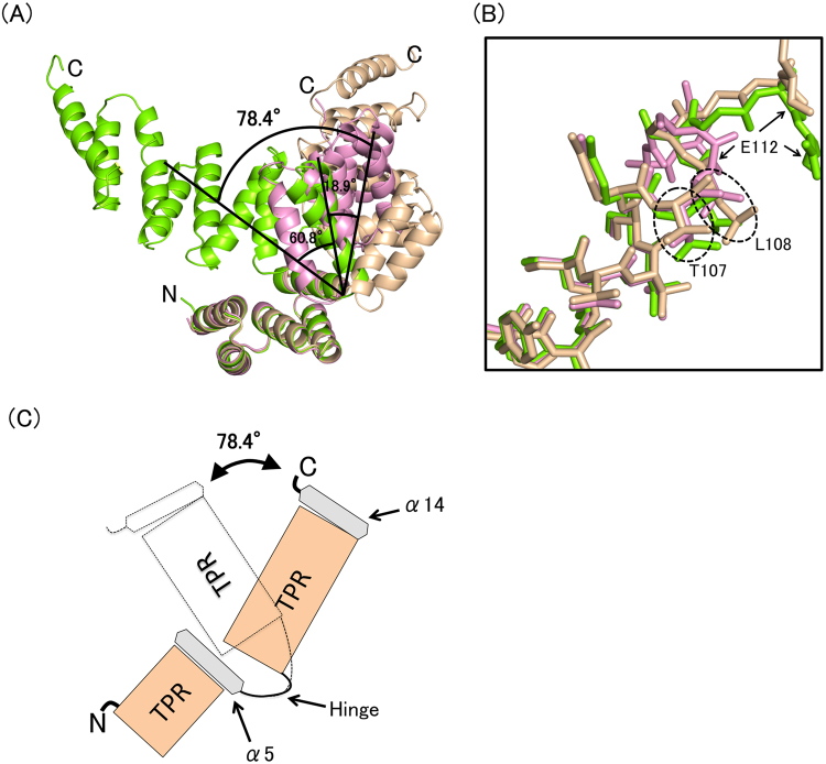
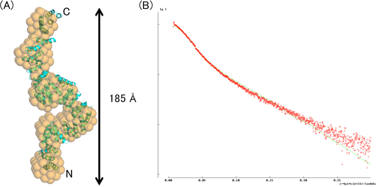
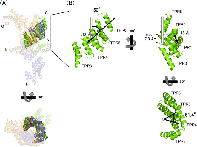
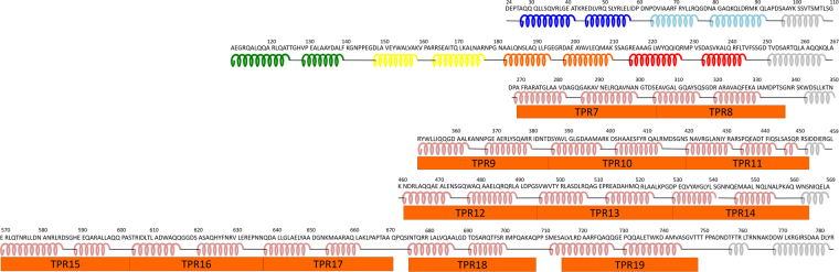
Bacterial cellulose (BC) is synthesized and exported through the cell membrane via a large protein complex (terminal complex) that consists of three or four subunits. BcsC is a little-studied subunit considered to export BC to the extracellular matrix. It is predicted to have two domains: a tetratrico peptide repeat (TPR) domain and a β-barrelled outer membrane domain. Here we report the crystal structure of the N-terminal part of BcsC-TPR domain (Asp24-Arg272) derived from Enterobacter CJF-002. Unlike most TPR-containing proteins which have continuous TPR motifs, this structure has an extra α-helix between two clusters of TPR motifs. Five independent molecules in the crystal had three different conformations that varied at the hinge of the inserted α-helix. Such structural feature indicates that the inserted α-helix confers flexibility to the chain and changes the direction of the TPR super-helix, which was also suggested by structural analysis of BcsC-TPR (Asp24-Leu664) in solution by size exclusion chromatography-small-angle X-ray scattering. The flexibility at the α-helical hinge may play important role for exporting glucan chains.


Conflict of interest statement

The authors declare that they have no competing interests.

Figures

Figure 1
Figure 1
The structure of BcsC-TPR(N6). (A) BcsC-TPR(N6) is composed of six TPR motifs (colored blue, light blue, green, yellow, orange, and red) and two unpaired α-helices (gray). (B) Schematic diagram of the secondary structure of BcsC-TPR(N6). The boxes indicate α-helices and the lines indicate turn parts. The color scheme is the same as (A).
Figure 2
Figure 2
The conformations of BcsC-TPR(N6). (A) Stereo view of five structures of BcsC-TPR(N6) in an asymmetric unit. (B) Five structures of BcsC-TPR(N6) formed three conformations (type 1, 2, and 3). BcsC-TPR(N6)s are shown in different colors (chain A: Green, chain B: Blue, chain C: wheat, chain D: orange, chain E: pink).
Figure 3
Figure 3
Structural comparison of BcsC-TPR(N6)s. (A) Superposition of three type chains on α1–α5. Black angles indicate the angle of the Cα of Ala219 (Chain A) and Leu108 (Chain A), Leu108 (Chain A) and Ala219 (Chain D). (B) Zoomed in and shown in stick format around the turn of inserted region. (C) Schematic image of the flexible region.
Figure 4
Figure 4
The structural analysis of BcsC-TPR in solution by SEC-SAXS. (A) The SAXS envelope (orange) and fitted structure of BcsC-TPR (cyan). Orange spheres indicate the SAXS envelope calculated from random dummy atoms. Cyan cartoon shows BcsC-TPR super-helices modeled by fitting a poly-Ala structure of BcsC-TPR(N6) as the TPR unit on the SAXS envelope. (B) Comparison between the calculated one-dimensional scattering intensity data (green line) of BcsC-TPR model and the measured one-dimensional scattering intensity data of SEC-SAXS (red dots).
Figure 5
Figure 5
Structural comparison of BcsC-TPR(N6)s. (A) Comparing TPR super-helices of different proteins. Green, BcsC-TPR(N6); blue, OGT; orange, IFIT5. Opaque cartoons indicate the TPR super-helices that are superposed. TPR super-helices are almost same structure. (B) The typical parameters of TPR super-helix. Green, SH2 of BcsC-TPR(N6); white stick, axis of super-helix.
Figure 6
Figure 6
Secondary structure and TPR motifs of BcsC-TPR domain (Asp24–Tyr783). The secondary structure elements are represented by coil (α-helix) and bar (loop), while the TPR motif predicted by TPRpred is represented by orange boxes. The secondary structure of the first two rows (BcsC-TPR(N6): Asp24–Ala267) are based on the crystal structure (colors are same as Fig. 1), and the other regions are based on the secondary structure prediction program (the PSIPRED server).

References

    1. Römling, U. & Galperin, M. Y. Bacterial cellulose biosynthesis: diversity of operons, subunits, products, and functions. Trends Microbiol. doi:10.1016/j.tim.2015.05.005 (2015). - PMC - PubMed
    1. Saxena IM, Brown RM. Cellulose biosynthesis: Current views and evolving concepts. Ann. Bot. 2005;96:9–21. doi: 10.1093/aob/mci155. - DOI - PMC - PubMed
    1. Ross P, Mayer R, Benziman M. Cellulose biosynthesis and function in bacteria. Microbiol. Rev. 1991;55:35–58. - PMC - PubMed
    1. Doblin MS. Cellulose Biosynthesis in Plants: from Genes to Rosettes. Plant Cell Physiol. 2002;43:1407–1420. doi: 10.1093/pcp/pcf164. - DOI - PubMed
    1. Kimura S, Kondo T. Recent progress in cellulose biosynthesis. J. Plant Res. 2002;115:297–302. doi: 10.1007/s10265-002-0037-7. - DOI - PubMed

Publication types

LinkOut - more resources