. 2017 Oct 12;7(1):13051.
doi: 10.1038/s41598-017-13442-9.

Genetic and Epigenetic Profiling Reveals EZH2-mediated Down Regulation of OCT-4 Involves NR2F2 during Cardiac Differentiation of Human Embryonic Stem Cells

Affiliations

Genetic and Epigenetic Profiling Reveals EZH2-mediated Down Regulation of OCT-4 Involves NR2F2 during Cardiac Differentiation of Human Embryonic Stem Cells

Varsha Pursani et al. Sci Rep. .

Abstract

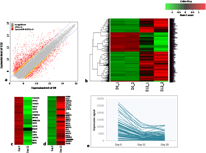
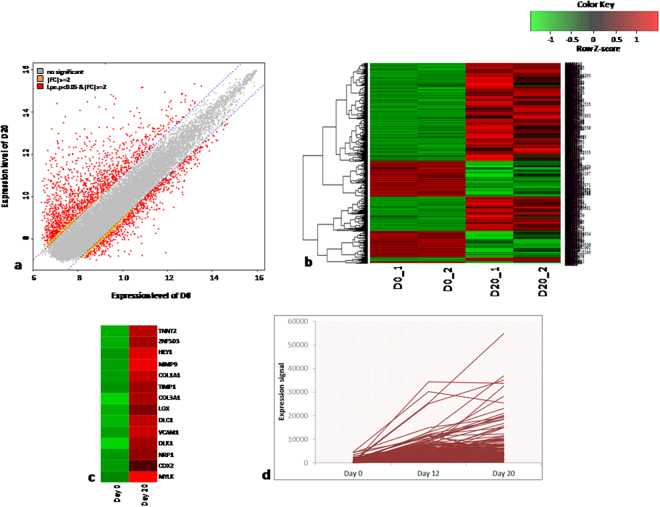
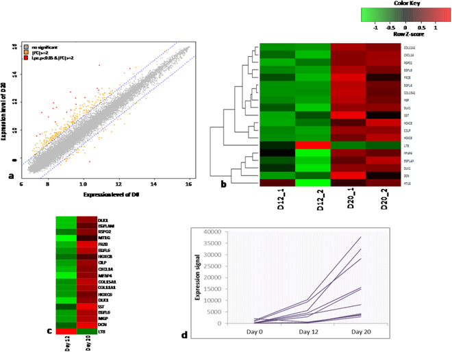
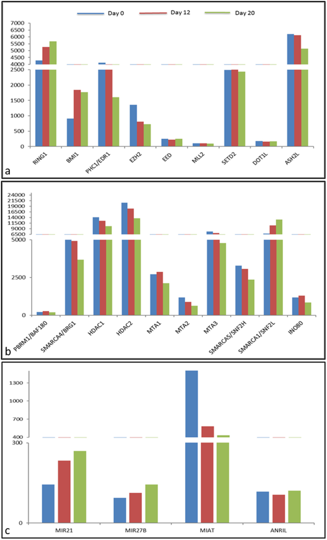
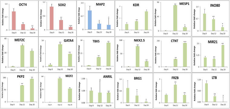
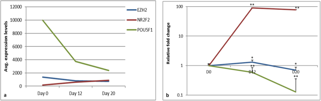
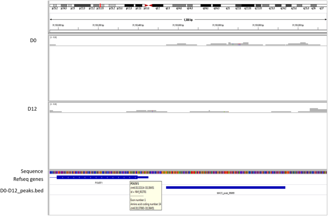
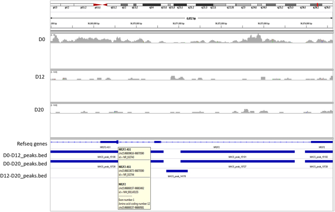
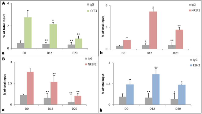
Human embryonic (hES) stem cells are widely used as an in vitro model to understand global genetic and epigenetic changes that occur during early embryonic development. In-house derived hES cells (KIND1) were subjected to directed differentiation into cardiovascular progenitors (D12) and beating cardiomyocytes (D20). Transcriptome profiling of undifferentiated (D0) and differentiated (D12 and 20) cells was undertaken by microarray analysis. ChIP and sequential ChIP were employed to study role of transcription factor NR2F2 during hES cells differentiation. Microarray profiling showed that an alteration of about 1400 and 1900 transcripts occurred on D12 and D20 respectively compared to D0 whereas only 19 genes were altered between D12 and D20. This was found associated with corresponding expression pattern of chromatin remodelers, histone modifiers, miRNAs and lncRNAs marking the formation of progenitors and cardiomyocytes on D12 and D20 respectively. ChIP sequencing and sequential ChIP revealed the binding of NR2F2 with polycomb group member EZH2 and pluripotent factor OCT4 indicating its crucial involvement in cardiac differentiation. The study provides a detailed insight into genetic and epigenetic changes associated with hES cells differentiation into cardiac cells and a role for NR2F2 is deciphered for the first time to down-regulate OCT-4 via EZH2 during cardiac differentiation.


Conflict of interest statement

The authors declare that they have no competing interests.

Figures

Figure 1
Figure 1
Differential gene expression profiling of D0 vs D12. (a) Plot of expression levels between D0 and D12. (b) Significant hierarchical clustering to show the clustering of the 2 biological replicates. (c) Heat map for the significant genes expressed on D0 and down regulated at D12. (d) Heat map for the significant genes not expressed at D0 and up regulated at D12. (e) Graphical plot for the expression levels of genes differentially expressed during conversion of D0 to D12 cells.
Figure 2
Figure 2
Differential gene expression profiling of D0 vs D20. (a) Plot of expression levels between D0 and D20. (b) Significant hierarchical clustering to show the clustering of the 2 biological replicates. (c) Heat map for the significant genes not expressed at D0 and up regulated at D20. (d) Graphical plot for the expression levels of genes differentially expressed during conversion of cells at D0 to D20.
Figure 3
Figure 3
Differential gene expression profiling of D12 vs D20. (a) Plot of expression levels between D12 and D20. (b) Significant hierarchical clustering to show the clustering of the 2 biological replicates. (c) Heat map for the significant genes not expressed at D12 and up regulated at D20. (e) Graphical plot for the expression levels of genes differentially expressed during conversion of cells at D12 to D20.
Figure 4
Figure 4
Expression of chromatin modifiers. Epigenetic analysis to identify the upregulated modifiers including histone modifiers (a), Chromatin remodelers (b) and non-coding RNAs (c) during different levels of cardiac differentiation.
Figure 5
Figure 5
Validation of Microarray data by qRTPCR. qRT-PCR validation for the crucial genes like OCT4, SOX2 (pluripotency) (red), MAP2 (ectodermal) (blue), KDR2, MESP1, MEF2C, GATA4, TBX5, NKX2.5, CTNT, PKP2, FRZB (mesodermal- cardiac), LTB, INO80, BRG1, MIR21 and ANRIL (epigenetic machinery) (green) in the D0, D12 and D20 cells profiled by microarray. Error bars represent ± SEM, statistical significance represented as *(P < 0.5), **(P < 0.01), ***(P < 0.001).
Figure 6
Figure 6
Expression pattern of EZH2, NR2F2 and OCT4. Relative expression levels of EZH2 (blue), NR2F2 (red) and OCT4 (green) by microarray (a) and qRT-PCR (b) during differentiation. Error bars represent ± SEM, statistical significance represented as *(P < 0.5), **(P < 0.01), ***(P < 0.001).
Figure 7
Figure 7
Distribution of H3K27me3 mark on OCT4. Binding profile of H3K27me3 mark on the OCT4 gene during cardiac differentiation generated by IGV genome browser mapped to human hg 19 genome.
Figure 8
Figure 8
Distribution of H3K27me3 mark on NR2F2. Occupancy of H3K27me3 mark on the NR2F2 locus at days 0, 12 and 20 generated by IGV gene browser mapped to human hg 19 genome.
Figure 9
Figure 9
ChIP and sequential ChIP. (A) Chromatin immuno-precipitation with anti-EZH2 followed by qPCR with NR2F2 (a) and OCT4 (b). The binding analysis shows that EZH2 binding increases on the NR2F2 promoter at D12 that later decrease at D20 while on the OCT4 promoter the EZH2 binding is maintained with differentiation (B) EZH2-OCT4 sequential ChIP followed by qPCR for genes EZH2 (a) and NR2F2 (b). The experiment confirms the c0-binding of EZH2 and OCT4 on the NR2F2 promoter at D12 that decreases slightly at D20. Values in both ChIP and sequential ChIP represent the average and standard deviation of two independent experiments and were calculated as percentage of total input. Error bars represent ± SEM, statistical significance represented as *(P < 0.5), **(P < 0.01), ***(P < 0.001).

References

    1. Thomson JA, et al. Embryonic stem cell lines derived from human blastocysts. Science. 1998;282:1145–7. doi: 10.1126/science.282.5391.1145. - DOI - PubMed
    1. Kehat I, et al. Human embryonic stem cells can differentiate into myocytes with structural and functional properties of cardiomyocytes. J Clin Invest. 2001;108:407–14. doi: 10.1172/JCI200112131. - DOI - PMC - PubMed
    1. Zhu R, et al. Physical developmental cues for the maturation of human pluripotent stem cell-derived cardiomyocytes. Stem Cell Res Ther. 2014;5:117. doi: 10.1186/scrt507. - DOI - PMC - PubMed
    1. Christophersen NS, Helin K. Epigenetic control of embryonic stem cell fate. J Exp Med. 2010;207:2287–95. doi: 10.1084/jem.20101438. - DOI - PMC - PubMed
    1. Surani MA, Hayashi K, Hajkova P. Genetic and Epigenetic Regulators of Pluripotency. Cell. 2007;128:747–62. doi: 10.1016/j.cell.2007.02.010. - DOI - PubMed

Publication types

MeSH terms

Substances