. 2017 Oct 12;7(1):13065.
doi: 10.1038/s41598-017-13588-6.

Mechanisms involved in the triggering of neutrophil extracellular traps (NETs) by Candida glabrata during planktonic and biofilm growth

Affiliations

Mechanisms involved in the triggering of neutrophil extracellular traps (NETs) by Candida glabrata during planktonic and biofilm growth

Chad J Johnson et al. Sci Rep. .

Abstract

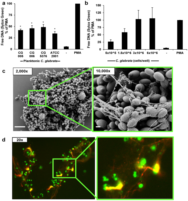
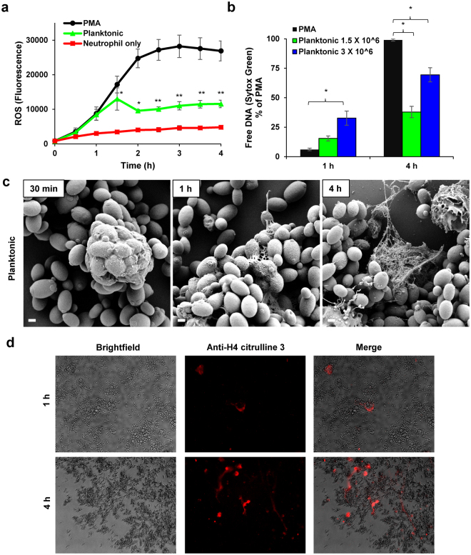
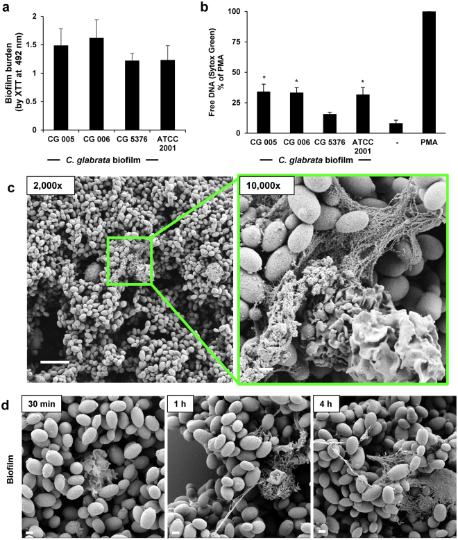
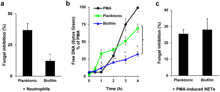
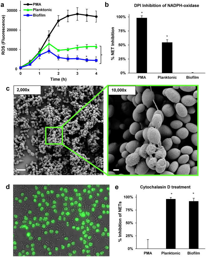
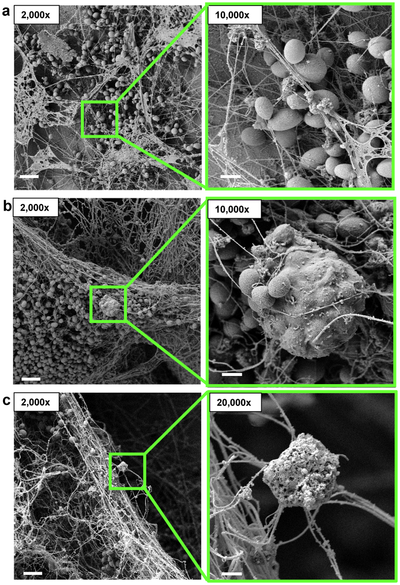
Candida spp. adhere to medical devices, such as catheters, forming drug-tolerant biofilms that resist killing by the immune system. Little is known about how C. glabrata, an emerging pathogen, resists attack by phagocytes. Here we show that upon encounter with planktonic (non-biofilm) C. glabrata, human neutrophils initially phagocytose the yeast and subsequently release neutrophil extracellular traps (NETs), complexes of DNA, histones, and proteins capable of inhibiting fungal growth and dissemination. When exposed to C. glabrata biofilms, neutrophils also release NETs, but significantly fewer than in response to planktonic cells. Impaired killing of biofilm parallels the decrease in NET production. Compared to biofilm, neutrophils generate higher levels of reactive oxygen species (ROS) when presented with planktonic organisms, and pharmacologic inhibition of NADPH-oxidase partially impairs NET production. In contrast, inhibition of phagocytosis nearly completely blocks NET release to both biofilm and planktonic organisms. Imaging of the host response to C. glabrata in a rat vascular model of infection supports a role for NET release in vivo. Taken together, these findings show that C. glabrata triggers NET release. The diminished NET response to C. glabrata biofilms likely contributes to the resilience of these structured communities to host defenses.


Conflict of interest statement

The authors declare that they have no competing interests.

Figures

Figure 1
Figure 1
Planktonic C. glabrata induce release of NETs. (a) Human neutrophils were exposed to four strains of planktonic C. glabrata (1.5 × 106 cells/well) for 4 h. NET release was estimated by Sytox Green detection of free DNA. Results were normalized for the positive control, PMA, and data from 5 experiments performed in triplicate were combined. Neutrophil responses to Candida were analyzed by ANOVA with pairwise comparison to the untreated neutrophil control, *P < 0.05, SEM shown. (b) Various concentrations of planktonic C. glabrata (CG 006) were co-cultured with human neutrophils for 4 h and NET release was estimated using Sytox Green. Results were normalized for the positive control, PMA, and data from 5 experiments performed in triplicate were combined, SEM shown. (c) Neutrophil interactions with planktonic C. glabrata at 4 h were imaged with scanning electron microscopy. Measurement bars represent 10 µm and 1 µm for 2,000x and 10,000x images, respectively. (d) Following co-culture with C. glabrata for 4 h, the neutrophil response was visualized by immunofluorescence using an anti-citrullinated H4 antibody (red) and Sytox Green staining of DNA (green).
Figure 2
Figure 2
Mechanism of NET release to planktonic C. glabrata. (a) Production of ROS in response to C. glabrata (CG 006 at 3 × 106 cells/well) was measured by fluorescence after neutrophils were pre-stained with oxidative stress indicator CM-DCF and co-cultured with C. glabrata over 4 h. The mean and SEM of 4 experiments performed in triplicate on 4 occasions are shown. Data for each time point were analyzed by ANOVA (*P < 0.05), with pairwise comparisons using the Holm-Sidak method (**P < 0.05 for C. glabrata v. PMA). (b) Neutrophils were co-cultured with planktonic C. glabrata for various time points and NET production was estimated by Sytox Green. Results were normalized to the positive control, PMA, and data from 4 experiments performed in triplicate were combined, SEM shown. Data for each time point were analyzed by ANOVA (*P < 0.05 for pairwise comparison by Holm-Sidak). (c) The neutrophil response to planktonic C. glabrata was imaged with scanning electron microscopy at various time points over 4 h. Measurement bars represent 1 µm for 10,000x images. (d) Following co-culture with C. glabrata for 1 h or 4 h, samples were immunolabeled with an anti-citrullinated H4 antibody and a fluorescently labeled (DyLight) secondary antibody and examined with brightfield microscopy and fluorescent (565/620 nm) microscopy.
Figure 3
Figure 3
Biofilms formed by C. glabrata elicit NET release. (a) The biofilm-forming capacity of C. glabrata isolates was estimated by XTT assay after 24 h of growth. Assays were performed in triplicate on 3 days and representative data is shown with SD. (b) C. glabrata biofilms (24 h) were co-cultured with human neutrophils and NET release was estimated by Sytox Green detection of free DNA. Results were normalized to the positive control, PMA, and data from 5 experiments performed in triplicate were combined. Neutrophil responses to Candida were analyzed by ANOVA with pairwise comparison to the untreated neutrophil control, *P < 0.05, SEM shown. (c) Neutrophil interactions with C. glabrata at 4 h were imaged with scanning electron microscopy. Measurement bars represent 10 µm and 1 µm for 2,000x and 10,000x images, respectively. (d) The neutrophil response to C. glabrata biofilms was imaged with scanning electron microscopy at various time points over 4 h. Measurement bars represent 1 µm for 10,000x images.
Figure 4
Figure 4
Comparison of neutrophil responses to C. glabrata during biofilm and planktonic growth. (a) Planktonic and biofilm C. glabrata were co-cultured with human neutrophils at an effector:target ratio of 1:2 for 4 h and fungal inhibition was estimated by an XTT assay. Results were normalized to the no neutrophil controls, and data from 3 experiments performed in triplicate were combined. Statistical significance was determined using a two-tailed Student’s t-test assuming unequal variances, *P < 0.05, SEM shown. (b) Neutrophils were co-cultured with C. glabrata for 4 h and NET release was estimated by Sytox Green staining of free DNA at various time points. Results were normalized for the positive control, PMA, and data from 4 experiments performed in triplicate were combined. The statistical significance for NET release to biofilm and planktonic C. glabrata were analyzed for each time point using a two-tailed Student’s t-test assuming unequal variances, *P < 0.05, SEM shown. (c) NETs were induced by incubation with PMA for 1.5 h prior to addition to C. glabrata. After 10 min, fungal inhibition was estimated by an XTT assay. Results were normalized to the no neutrophil controls, and data from 4 experiments performed in triplicate were combined. Statistical significance was determined using a two-tailed Student’s t-test assuming unequal variances, *P < 0.05, SEM shown.
Figure 5
Figure 5
Mechanism of NET induction by planktonic and biofilm C. glabrata. (a) Production of ROS in response to C. glabrata was measured by fluorescence after neutrophils were pre-stained with oxidative stress indicator CM-H2DCFDA and co-cultured with C. glabrata over 4 h. The mean and SEM of 4 experiments performed in triplicate is shown. The statistical significance neutrophil production of ROS in response to biofilm and planktonic C. glabrata were calculated for each time point using a two-tailed Student’s t-test assuming unequal variances, *P < 0.05, SEM shown. (b) Neutrophils were treated with DPI to inhibit NADPH-oxidase and the release of NETs in response to C. glabrata was measured by Sytox Green. The percent of the total free DNA (untreated) reduced by DPI-treatment for each condition is shown. Data represent 5 experiments performed in triplicate. Statistical significance was determined using a Student’s t-test compared to no inhibition, *P < 0.05, SEM shown. (c) Neutrophil-C. glabrata interactions after 4 h were imaged with scanning electron microscopy. (d) Calcein AM-labeled neutrophils (green) were treated with cytochalasin D to inhibit phagocytosis and added to planktonic C. glabrata. Neutrophil interactions were imaged at 1 h (40x). (e) Neutrophils were treated with cytochalasin D to inhibit phagocytosis and the release of NETs in response to C. glabrata was measured by Sytox Green. The percent of the total free DNA (untreated) reduced by DPI treatment for each condition is shown. Data represent 5 experiments performed in triplicate. Statistical significance was determined using a Student’s t-test compared to no inhibition, *P < 0.05, SEM shown.
Figure 6
Figure 6
C. glabrata appears to induce the formation of NETs in vivo. (a,b,c) C. glabrata was inoculated in the lumen of rat jugular catheters. After 48 h, catheters were harvested and host-fungal interactions on the luminal catheter surface were observed by scanning electron microscopy. Measurement bars represent 10 µm and 1 µm for 2,000x and 10,000x images, respectively.

Similar articles

Cited by

References

    1. Pfaller MA, Diekema DJ. Epidemiology of invasive candidiasis: a persistent public health problem. Clin Microbiol Rev. 2007;20:133–163. doi: 10.1128/CMR.00029-06. - DOI - PMC - PubMed
    1. Andes DR, et al. Impact of treatment strategy on outcomes in patients with candidemia and other forms of invasive candidiasis: a patient-level quantitative review of randomized trials. Clin Infect Dis. 2012;54:1110–1122. doi: 10.1093/cid/cis021. - DOI - PubMed
    1. Horn DL, et al. Epidemiology and outcomes of candidemia in 2019 patients: data from the prospective antifungal therapy alliance registry. Clin Infect Dis. 2009;48:1695–1703. doi: 10.1086/599039. - DOI - PubMed
    1. Pappas PG, et al. Clinical Practice Guideline for the Management of Candidiasis: 2016 Update by the Infectious Diseases Society of America. Clin Infect Dis. 2016;62:e1–e50. doi: 10.1093/cid/civ1194. - DOI - PMC - PubMed
    1. Donlan RM. Biofilm formation: a clinically relevant microbiological process. Clin Infect Dis. 2001;33:1387–1392. doi: 10.1086/322972. - DOI - PubMed

Publication types

Substances