Review
. 2017 Nov;17(11):659-675.
doi: 10.1038/nrc.2017.83. Epub 2017 Oct 13.

Engineering and physical sciences in oncology: challenges and opportunities

Affiliations
Review

Engineering and physical sciences in oncology: challenges and opportunities

Michael J Mitchell et al. Nat Rev Cancer. 2017 Nov.

Erratum in

Abstract

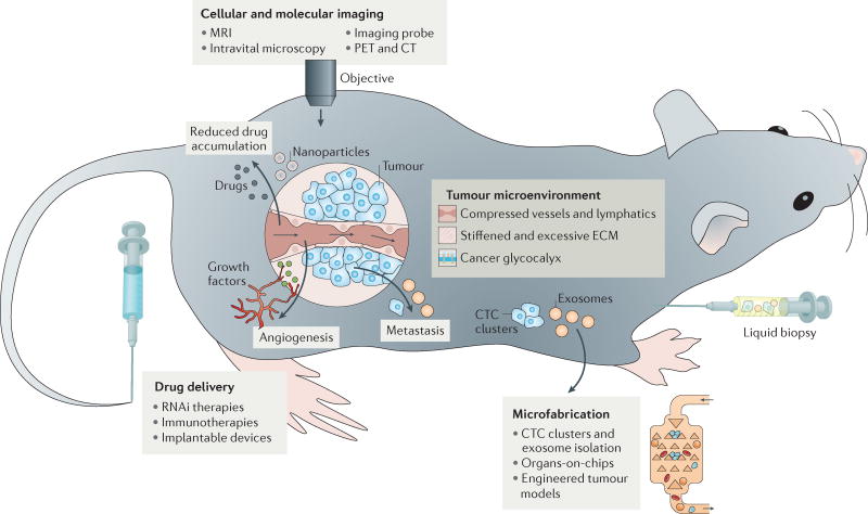
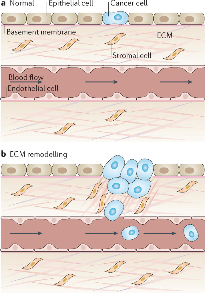
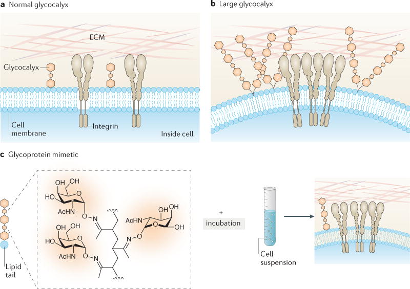
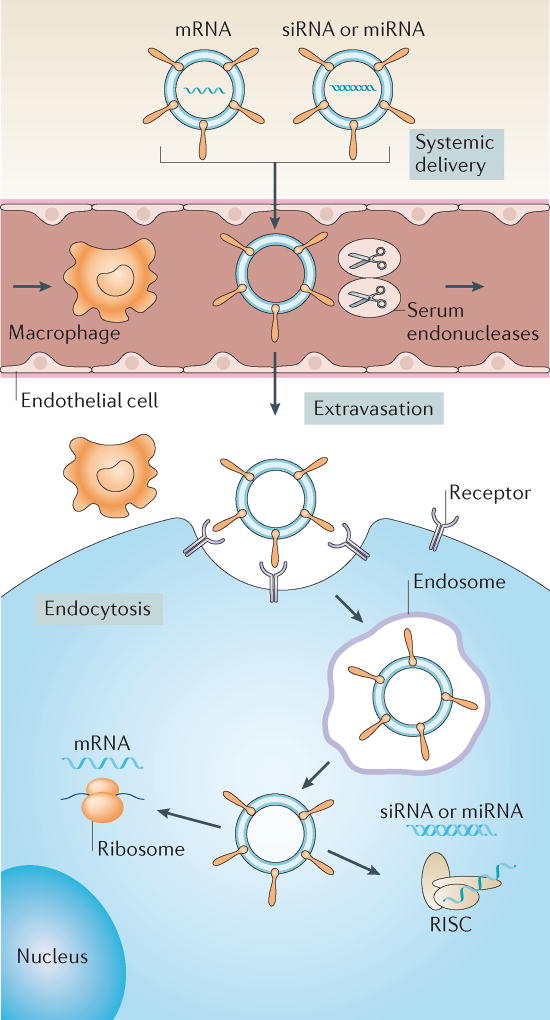
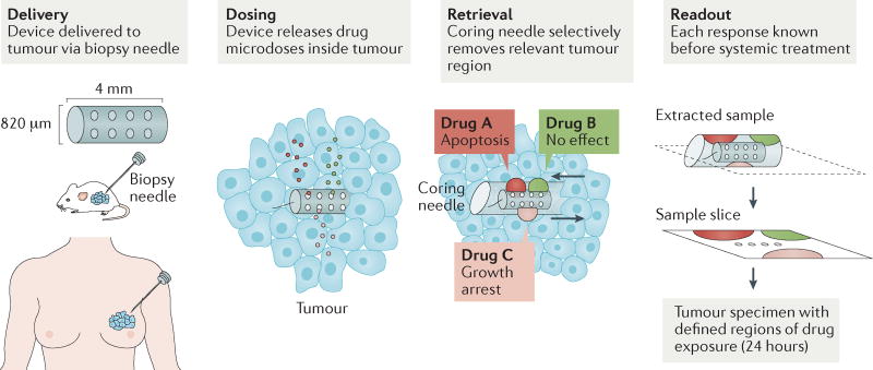
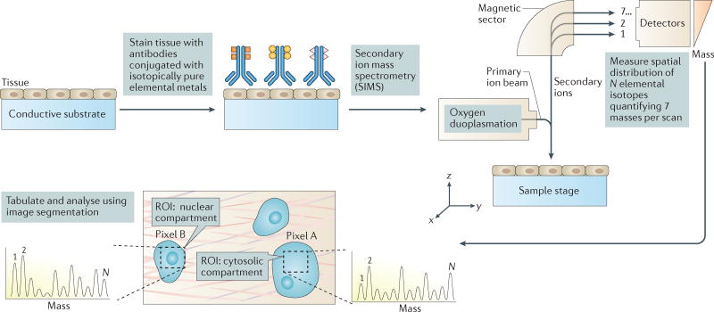
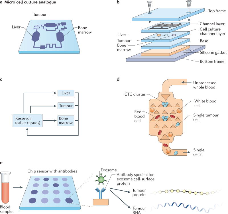
The principles of engineering and physics have been applied to oncology for nearly 50 years. Engineers and physical scientists have made contributions to all aspects of cancer biology, from quantitative understanding of tumour growth and progression to improved detection and treatment of cancer. Many early efforts focused on experimental and computational modelling of drug distribution, cell cycle kinetics and tumour growth dynamics. In the past decade, we have witnessed exponential growth at the interface of engineering, physics and oncology that has been fuelled by advances in fields including materials science, microfabrication, nanomedicine, microfluidics, imaging, and catalysed by new programmes at the National Institutes of Health (NIH), including the National Institute of Biomedical Imaging and Bioengineering (NIBIB), Physical Sciences in Oncology, and the National Cancer Institute (NCI) Alliance for Nanotechnology. Here, we review the advances made at the interface of engineering and physical sciences and oncology in four important areas: the physical microenvironment of the tumour and technological advances in drug delivery; cellular and molecular imaging; and microfluidics and microfabrication. We discussthe research advances, opportunities and challenges for integrating engineering and physical sciences with oncology to develop new methods to study, detect and treat cancer, and we also describe the future outlook for these emerging areas.


Conflict of interest statement

Competing interests statement

The authors declare no competing interests.

Figures

Figure 1
Figure 1. An overview of engineering and physical sciences in oncology
Physical abnormalities of the tumour microenvironment (TME) have been identified using tools and concepts from engineering and the physical sciences. These include blood vessel and lymphatic compression, stiffened and excessive extracellular matrix (ECM), and the cancer cell glycocalyx. Collapsed blood vessels and increased solid stresses lead to reduced accumulation and limited delivery of drugs to tumour tissues. Steep pressure gradients in the periphery push fluid leaking from blood vessels located in the tumour margin into the surrounding normal tissues, facilitating the transport of growth factors and cancer cells into normal tissue and thus fuelling tumour growth, angiogenesis and metastasis. Pressure gradients also reduce the retention time of drugs and inhibit their homogeneous distribution inside the tumour. Advances in imaging, drug delivery and microfabrication have all been used to detect, manipulate and therapeutically target various aspects of this microenvironment. CT, computed tomography; CTC, circulating tumour cell; MRI, magnetic resonance imaging; PET, positron emission tomography.
Figure 2
Figure 2. Extracellular matrix stiffening promotes cancer progression
a | Under homeostatic conditions, the extracellular matrix (ECM) maintains tissue integrity and blocks rare tumour-prone cells from malignant progression by maintaining an overall healthy microenvironment. b | Under pathological conditions, ECM remodelling leads to collagen fibre alignment, bundling, and stiffening, which in turn alter interactions between the matrix and stromal and tumour cells to enhance pro-angiogenic secretion from a range of cells in the microenvironment as well as the migration of cancer cells. This process consequently promotes both the invasion of tumour cells from the primary site into the circulation and the recruitment of endothelial cells for vascularization of the tumour to initiate tumour growth, invasion into the surrounding stroma and, finally, metastasis.
Figure 3
Figure 3. Role of the cancer cell glycocalyx in cancer progression
a | Normal cells with a short glycocalyx have a uniform distribution of glycoproteins and adhesion molecules (integrins) across the cell membrane, which is close to the surrounding extracellular matrix (ECM). b | Cells with a larger glycocalyx, such as tumour cells, exhibit extended gaps between the membrane and ECM, clustering of integrins, the exclusion of glycopolymers from regions of integrin adhesion, and membrane bending. These physical effects can alter cell signalling and promote tumour survival. c | Engineering the cancer cell glycocalyx via incorporation of synthetic glycoprotein mimetics with lipid insertion domains into living cell membranes. The glycopolymers consist of a long-chain polymer backbone, pendant glycan chains mimicking natural mucin O-glycans, a phospholipid insertion domain and a fluorophore for imaging incorporation into cells. The approach enables synthetic mucin glycoprotein mimetics of a range of lengths to be rapidly incorporated into plasma membranes, where they project perpendicular to the cell surface. Synthetic glycoprotein mimetics have been used to study how the physical properties of the glycocalyx coating regulate cell survival during tumour invasion. Parts a and b are from REF. , Macmillan Publishers Limited.
Figure 4
Figure 4. Drug delivery vehicles to enhance cancer immunotherapy
a | A nanoparticle library is engineered to have varying surface charge in the absence of targeting ligands. Nanoparticle charge is altered by tailoring the ratio of the lipid delivery material to the amount of encapsulated RNA. These materials are then screened in mice for efficient delivery and transfection of dendritic cells in the spleen and other lymphoid organs. The top candidate with a slightly negative charge is delivered into mice via a nanoparticle RNA vaccine to target precursor dendritic cells, which causes them to develop into mature antigen-presenting dendritic cells that migrate to the T cells in lymph nodes. b | Uptake by plasmacytoid dendritic cells promotes secretion of an initial wave of interferon-α (IFNα) production that helps to prime initial T cell activation in lymph nodes. c | Mature dendritic cells express tumour antigens derived from RNA, adjuvant or antigen in delivery vehicles and present them to T cells in lymph nodes. d | Uptake of delivery vehicles by macrophages leads to a second wave of IFNα release, fully priming T cells against specific antigens. Primed T cells then migrate to tumour sites, attacking and killing tumour cells. Figure from REF. , Macmillan Publishers Limited.
Figure 5
Figure 5. Non-viral delivery vectors for RNA-based therapies
Various non-viral vectors, such as nanoparticles, can be used to deliver mRNA, small interfering RNA (siRNA) or microRNA (miRNA) therapeutics to target cells in vivo. These vectors prevent degradation of nucleic acids by serum endonucleases and evade immune detection. For effective delivery, these vehicles need to avoid renal clearance from the circulation and prevent nonspecific interactions with cells and proteins. When delivered intravenously, these vectors need to (i) extravasate from the bloodstream to reach target tissues, owing to nanoparticle characteristics and/or targeting ligands, (ii) enter target cells via the plasma membrane and (iii) induce endosomal escape into the cytosol. siRNA and miRNA must be loaded into the RNA-induced silencing complex (RISC) to initiate RNAi, whereas mRNA binds to translational machinery for subsequent protein expression. Figure from REF. , Macmillan Publishers Limited.
Figure 6
Figure 6. Implantable drug delivery devices for simultaneous screening of many drugs in tumours
Implantable drug delivery devices were recently developed to enable in vivo drug sensitivity testing and biomarker analysis in patient tumours. One such device that can be implanted directly into tumours via biopsy needle is shown. It can be used to administer and subsequently evaluate the effects of up to 16 different drugs simultaneously via drug-releasing microwells. Three drugs released from microwells are depicted here for simplicity. The drugs diffuse from the microwells into confined regions of the tumour. The tumour tissue is then biopsied using a second coring needle that retrieves the device itself and a small column of tissue adjacent to the device. This tissue contains regions exposed to the drugs and is used to evaluate drug effects, such as apoptosis or growth arrest. Another delivery device, the CIVO platform (not shown in this figure), microinjects up to six different drugs into tumours as they are being withdrawn, leaving a 6 mm track of both the drug and inert tracking dye. Tumour cell death in response to drugs is assessed 24–72 hours after injection, via tumour resection. Evaluation of pharmacological and pharmacodynamic markers in these studies, such as cleaved caspase 3 as a marker of tumour cell apoptosis, demonstrated that device outputs were similar to the effects of systemic in vivo therapy. These devices offer a possible alternative to the traditional way of using cancer drugs that has become accepted practice for clinical trials and animal research. Both devices potentially offer a personalized system for assessing drug sensitivity in vivo and tailoring therapy accordingly. Additionally, both devices provide ease of testing several drug combinations directly within tumours, along with probing inter-tumour and intra-tumour heterogeneity in their response to drugs. Figure from REF. : Jonas, O. et al. An implantable microdevice to perform high-throughput in vivo drug sensitivity testing in tumors. Science Translational Medicine 7, 284ra57 (2015). Reprinted with permission from AAAS.
Figure 7
Figure 7. Multiplexed ion beam imaging for detecting as many as 100 targets simultaneously in tumour tissue samples
Cell and tissue samples are immobilized on a conductive substrate and subsequently stained with antibodies conjugated to unique, isotopically pure elemental metal reporters. Samples are then dried and loaded under vacuum for multiplexed ion beam imaging (MIBI) analysis, in which the surface is rasterized with an oxygen primary ion beam that sputters the antibody-specific metal reporters native to the sample surface as secondary ions. Metal-conjugated antibodies are quantified via replicate scans of the same field of view, and regions of interest (ROIs) demarcating nuclear and cytosolic compartments of cells within the sample are integrated, tabulated and categorized. From these expression data, composite images composed of pseudocoloured categorical features, and quantitative three-colour overlays are then constructed. MIBI is capable of detecting up to 100 unique isotope-labelled antibodies and has been used to analyse paraffin-embedded human breast cancer tissue samples stained simultaneously with ten isotope-labelled antibodies to detect features such as nuclear and cytosolic compartments of cells, providing new insights into disease pathogenesis for basic research and clinical diagnostics. N = number of unique elemental reporters. Figure from REF. , Macmillan Publishers Limited.
Figure 8
Figure 8. Microfluidics and microfabricated devices for ‘organs-on-chip’ tumour models and cancer diagnostics
a | A microfluidic device containing interconnected, 3D cell culture microchambers that mimic tissues (tumour, bone marrow, liver) to develop a multi-organ model that simulates absorption, metabolism and activity of chemotherapeutic agents. b | Schematic of the microfabrication of the three-chamber organs-on-chip that is linked via microfluidics. c | Flow diagram of the connections between the tumour, liver and bone marrow compartments of the microfluidic organs-on-chip model. Drugs are added into the culture medium, which is recirculated in a controlled manner through three inline chambers and an external reservoir, to mimic physiological blood flow rates and blood residence times in each organ. Mathematical models are also utilized for fitting to experimental toxicity measurements, and parameter optimization is used to mimic liver, tumour and bone marrow cytotoxicity in vivo. d | The Cluster-Chip device that captures circulating tumour cell (CTC) clusters from flowing unprocessed whole blood via microfabricated triangular micropillars, while single blood and tumour cells pass through the device. e | Exosomes can be efficiently captured from blood using a nano-plasmonic exosome sensor, an array of periodic nanoholes patterned in gold film. Exosomes are captured on the sensors via affinity ligands specific for protein markers characteristic of exosomes, such as CD63. Exosome binding to the array changes the local refractive index of the sensor to an extent proportional to the level of the target protein, and can be used to detect the concentration of exosomes as well the abundance of proteins on or within exosomes. As a result of this chip sensor technique, rare tumour proteins and RNA can then be extracted from exosomes for further analysis. Part a is from REF. , Macmillan Publishers Limited. Parts b and c are from REF. , Macmillan Publishers Limited. Part e is from REF. , Macmillan Publishers Limited.

References

    1. Jain RK, Martin JD, Stylianopoulos T. The role of mechanical forces in tumor growth and therapy. Annu. Rev. Biomed. Eng. 2014;16:321–346. Comprehensive review of the role of physical forces in tumour progression and therapy for those new to the fields of engineering and physical sciences in oncology. - PMC - PubMed
    1. Wirtz D, Konstantopoulos K, Searson PC. The physics of cancer: the role of physical interactions and mechanical forces in metastasis. Nat. Rev. Cancer. 2011;11:512–522. - PMC - PubMed
    1. Lu P, Weaver VM, Werb Z. The extracellular matrix: a dynamic niche in cancer progression. J. Cell Biol. 2012;196:395–406. - PMC - PubMed
    1. Tse JM, et al. Mechanical compression drives cancer cells toward invasive phenotype. Proc. Natl Acad. Sci. USA. 2012;109:911–916. - PMC - PubMed
    1. Jain RK. Antiangiogenesis strategies revisited: from starving tumors to alleviating hypoxia. Cancer Cell. 2014;26:605–622. - PMC - PubMed

Publication types