

Murine hepatocellular carcinoma derived stem cells reveal epithelial-to-mesenchymal plasticity
- PMID: 29026462
- PMCID: PMC5620425
- DOI: 10.4252/wjsc.v9.i9.159
Murine hepatocellular carcinoma derived stem cells reveal epithelial-to-mesenchymal plasticity
Abstract
Aim: To establish a model to enrich and characterize stem-like cells from murine normal liver and hepatocellular carcinoma (HCC) cell lines and to further investigate stem-like cell association with epithelial-to-mesenchymal transition (EMT).
Methods: In this study, we utilized a stem cell conditioned serum-free medium to enrich stem-like cells from mouse HCC and normal liver cell lines, Hepa 1-6 and AML12, respectively. We isolated the 3-dimensional spheres and assessed their stemness characteristics by evaluating the RNA levels of stemness genes and a cell surface stem cell marker by quantitative reverse transcriptase-PCR (qRT-PCR). Next, we examined the relationship between stem cells and EMT using qRT-PCR.
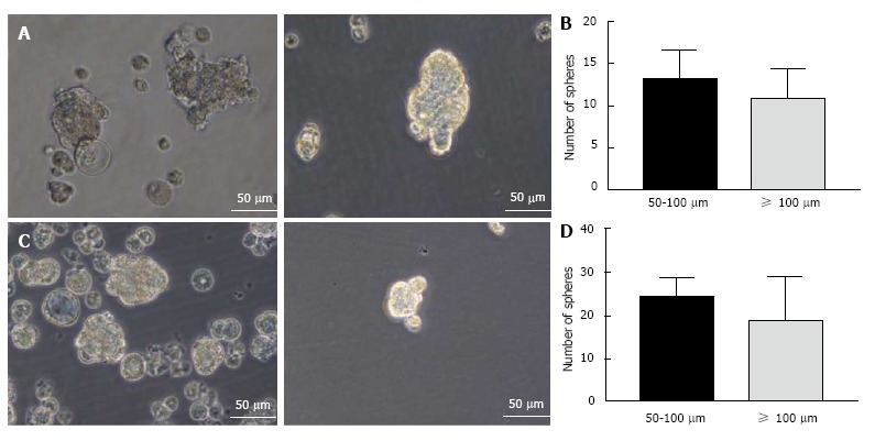
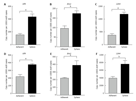
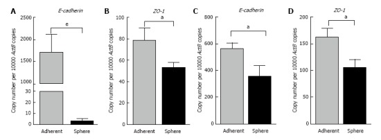
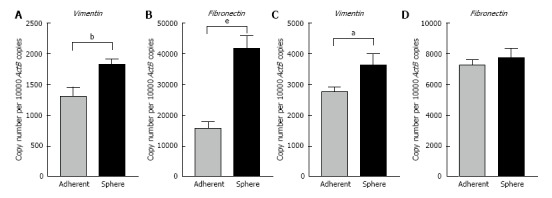
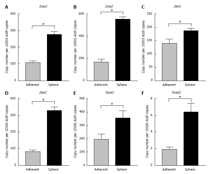
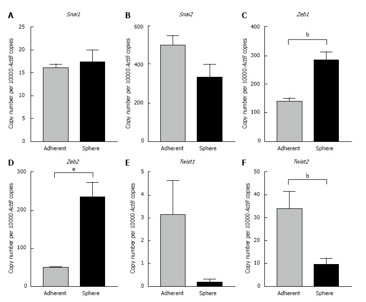
Results: Three-dimensional spheres were enriched by culturing murine HCC and normal hepatocyte cell lines in stem cell conditioned serum-free medium supplemented with epidermal growth factor, basic fibroblast growth factor and heparin sulfate. The 3-dimensional spheres had enhanced stemness markers such as Klf4 and Bmi1 and hepatic cancer stem cell (CSC) marker Cd44 compared to parental cells grown as adherent cultures. We report that epithelial markers E-cadherin and ZO-1 were downregulated, while mesenchymal markers Vimentin and Fibronectin were upregulated in 3-dimensional spheres. The 3-dimensional spheres also exhibited changes in expression of Snai, Zeb and Twist family of EMT transcription factors.
Conclusion: Our novel method successfully enriched stem-like cells which possessed an EMT phenotype. The isolation and characterization of murine hepatic CSCs could establish a precise target for the development of more effective therapies for HCC.
Keywords: AML12; Cancer initiating cells; Cancer stem cells; Cellular plasticity; Epithelial-to-mesenchymal transition; Epithelial-to-mesenchymal transition transcription factors; Hepa 1-6; Hepatocellular carcinoma.
Conflict of interest statement
Conflict-of-interest statement: The authors declare no conflict of interest. The views expressed in this article are those of the author and do not necessarily reflect the official policy or position of the Department of the Navy, Department of Defense, nor the United States Government. Brian J Morrison is a military service member. This work was prepared as part of his official duties. Title 17 U.S.C. §105 provides that Copyright protection under this title is not available for any work of the United States Government. Title 17 U.S.C. §101 defines a United States Government work as a work prepared by a military service member or employee of the United States Government as part of that person’s official duties.
Figures
Similar articles
-
Induction of epithelial-mesenchymal transition (EMT) and Gli1 expression in head and neck squamous cell carcinoma (HNSCC) spheroid cultures.Bosn J Basic Med Sci. 2018 Nov 7;18(4):336-346. doi: 10.17305/bjbms.2018.3243. Bosn J Basic Med Sci. 2018. PMID: 30172250 Free PMC article.
-
Identification of biomarkers associated with partial epithelial to mesenchymal transition in the secretome of slug over-expressing hepatocellular carcinoma cells.Cell Oncol (Dordr). 2018 Aug;41(4):439-453. doi: 10.1007/s13402-018-0384-6. Epub 2018 Jun 1. Cell Oncol (Dordr). 2018. PMID: 29858962
-
Epithelial to mesenchymal transition is associated with shorter disease-free survival in hepatocellular carcinoma.Ann Surg Oncol. 2014 Nov;21(12):3882-90. doi: 10.1245/s10434-014-3779-2. Epub 2014 May 15. Ann Surg Oncol. 2014. PMID: 24833103
-
Epithelial-to-mesenchymal plasticity of cancer stem cells: therapeutic targets in hepatocellular carcinoma.J Hematol Oncol. 2016 Aug 30;9(1):74. doi: 10.1186/s13045-016-0307-9. J Hematol Oncol. 2016. PMID: 27578206 Free PMC article. Review.
-
Communication Between Epithelial-Mesenchymal Plasticity and Cancer Stem Cells: New Insights Into Cancer Progression.Front Oncol. 2021 Apr 21;11:617597. doi: 10.3389/fonc.2021.617597. eCollection 2021. Front Oncol. 2021. PMID: 33968721 Free PMC article. Review.
Cited by
-
miRNA122a regulation of gene therapy vectors targeting hepatocellular cancer stem cells.Oncotarget. 2018 May 4;9(34):23577-23588. doi: 10.18632/oncotarget.25280. eCollection 2018 May 4. Oncotarget. 2018. PMID: 29805757 Free PMC article.
-
Tobacco smoke induced hepatic cancer stem cell-like properties through IL-33/p38 pathway.J Exp Clin Cancer Res. 2019 Jan 28;38(1):39. doi: 10.1186/s13046-019-1052-z. J Exp Clin Cancer Res. 2019. PMID: 30691509 Free PMC article.
-
MicroRNA199a-Based Post-transcriptional Detargeting of Gene Vectors for Hepatocellular Carcinoma.Mol Ther Nucleic Acids. 2018 Dec 7;13:78-88. doi: 10.1016/j.omtn.2018.08.016. Epub 2018 Aug 24. Mol Ther Nucleic Acids. 2018. PMID: 30245470 Free PMC article.
-
The role of proteasomes in tumorigenesis.Genes Dis. 2023 Aug 6;11(4):101070. doi: 10.1016/j.gendis.2023.06.037. eCollection 2024 Jul. Genes Dis. 2023. PMID: 38523673 Free PMC article. Review.
-
Immune checkpoint molecules are regulated by transforming growth factor (TGF)-β1-induced epithelial-to-mesenchymal transition in hepatocellular carcinoma.Int J Med Sci. 2021 Apr 22;18(12):2466-2479. doi: 10.7150/ijms.54239. eCollection 2021. Int J Med Sci. 2021. PMID: 34104078 Free PMC article.
References
-
- Thorgeirsson SS, Grisham JW. Molecular pathogenesis of human hepatocellular carcinoma. Nat Genet. 2002;31:339–346. - PubMed
-
- El-Serag HB, Rudolph KL. Hepatocellular carcinoma: epidemiology and molecular carcinogenesis. Gastroenterology. 2007;132:2557–2576. - PubMed
-
- Mir N, Jayachandran A, Dhungel B, Shrestha R, Steel JC. Epithelial-to-Mesenchymal Transition: a Mediator of Sorafenib Resistance in Advanced Hepatocellular Carcinoma. Curr Cancer Drug Targets. 2017 - PubMed
-
- Llovet JM, Ricci S, Mazzaferro V, Hilgard P, Gane E, Blanc JF, de Oliveira AC, Santoro A, Raoul JL, Forner A, et al. Sorafenib in advanced hepatocellular carcinoma. N Engl J Med. 2008;359:378–390. - PubMed
-
- Reya T, Morrison SJ, Clarke MF, Weissman IL. Stem cells, cancer, and cancer stem cells. Nature. 2001;414:105–111. - PubMed
LinkOut - more resources
Full Text Sources
Other Literature Sources
Miscellaneous
