Review
. 2017 Aug 31:6:1622.
doi: 10.12688/f1000research.11652.1. eCollection 2017.

Recent advances in pericentriolar material organization: ordered layers and scaffolding gels

Affiliations
Review

Recent advances in pericentriolar material organization: ordered layers and scaffolding gels

Andrew M Fry et al. F1000Res. .

Abstract

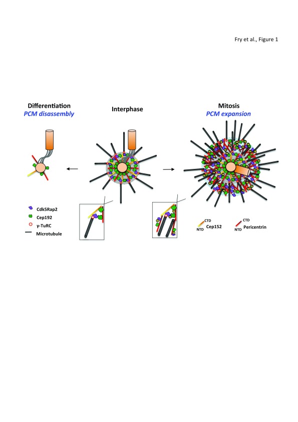
The centrosome is an unusual organelle that lacks a surrounding membrane, raising the question of what limits its size and shape. Moreover, while electron microscopy (EM) has provided a detailed view of centriole architecture, there has been limited understanding of how the second major component of centrosomes, the pericentriolar material (PCM), is organized. Here, we summarize exciting recent findings from super-resolution fluorescence imaging, structural biology, and biochemical reconstitution that together reveal the presence of ordered layers and complex gel-like scaffolds in the PCM. Moreover, we discuss how this is leading to a better understanding of the process of microtubule nucleation, how alterations in PCM size are regulated in cycling and differentiated cells, and why mutations in PCM components lead to specific human pathologies.

Keywords: centrosomes; mitosis; pericentriolar material (PCM).


Conflict of interest statement

Competing interests: The authors declare that they have no competing interests.No competing interests were disclosed.No competing interests were disclosed.No competing interests were disclosed.

Figures

Figure 1.
Figure 1.. Expansion and disassembly of the PCM upon mitotic progression and differentiation.
This schematic figure provides a simplified overview of PCM organization. During interphase (center panel), the two centrioles (orange cylinders) are connected by an inter-centriolar linker with the bulk of the PCM associated with the older (mother) centriole. The size and shape of the PCM is defined by the two filament proteins, pericentrin and Cep152, that extend radially from the centriole surface to generate a proximal layer (gray circle). This contains other proteins, such as Cdk5Rap2 and Cep192, which together create a branched matrix that provides binding sites for γ-TuRCs and their adaptor proteins. A second population of Cep192 is closely associated with proteins involved in centriole duplication at the centriole surface. In mitosis (right panel), PCM expansion results from the phosphorylation of multiple proteins, including pericentrin, Cdk5Rap2, and Cep192, by Plk1. This creates an outer expansive layer with gel-like properties (blue hatched circle) that is less well ordered but contains scaffolds that increase the microtubule nucleation capacity necessary for spindle assembly. Indeed, increased levels of these PCM proteins, together with additional centrosomally localized tubulin-binding proteins, such as chTOG and TPX2, may well allow microtubule nucleation to occur independently of γ-TuRCs (see inset). In contrast, in certain differentiated cells (left panel), disassembly of the proximal layer occurs with PCM proteins recruited to other non-centrosomal MTOCs. Note that the interphase centrosome is shown with two unduplicated centrioles typical of a G1 cell, whereas the mitotic centrosome contains a duplicated centriole pair that has also lost the inter-centriolar linker (mitotic cells have four centrioles, two in each spindle pole). CTD, C-terminal domain; NTD, N-terminal domain.

References

    1. Conduit PT, Wainman A, Raff JW: Centrosome function and assembly in animal cells. Nat Rev Mol Cell Biol. 2015;16(10):611–24. 10.1038/nrm4062 - DOI - PubMed
    1. Kollman JM, Merdes A, Mourey L, et al. : Microtubule nucleation by γ-tubulin complexes. Nat Rev Mol Cell Biol. 2011;12(11):709–21. 10.1038/nrm3209 - DOI - PMC - PubMed
    1. Gould RR, Borisy GG: The pericentriolar material in Chinese hamster ovary cells nucleates microtubule formation. J Cell Biol. 1977;73(3):601–15. 10.1083/jcb.73.3.601 - DOI - PMC - PubMed
    1. Mennella V, Agard DA, Huang B, et al. : Amorphous no more: subdiffraction view of the pericentriolar material architecture. Trends Cell Biol. 2014;24(3):188–97. 10.1016/j.tcb.2013.10.001 - DOI - PMC - PubMed
    1. Woodruff JB, Wueseke O, Hyman AA: Pericentriolar material structure and dynamics. Philos Trans R Soc Lond B Biol Sci. 2014;369(1650): pii: 20130459. 10.1098/rstb.2013.0459 - DOI - PMC - PubMed