

Synthesis and characterization of collagen/PLGA biodegradable skin scaffold fibers
- PMID: 29026645
- PMCID: PMC5633691
- DOI: 10.1093/rb/rbx026
Synthesis and characterization of collagen/PLGA biodegradable skin scaffold fibers
Abstract
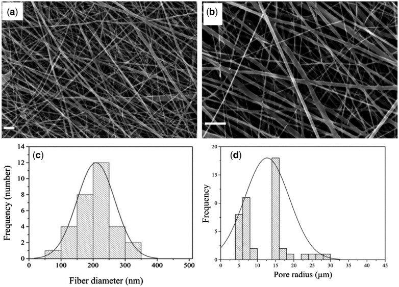
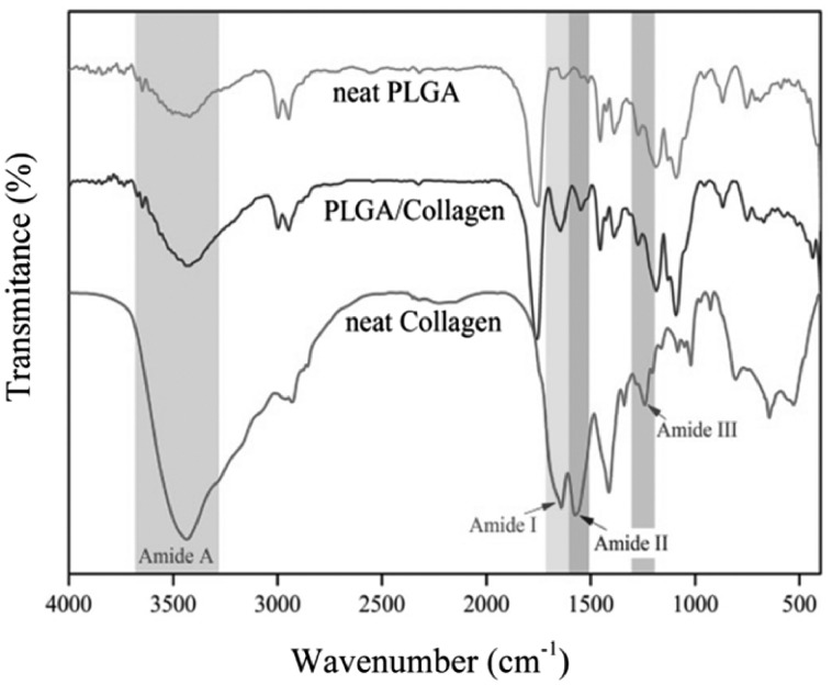
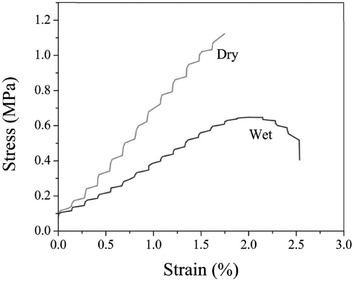
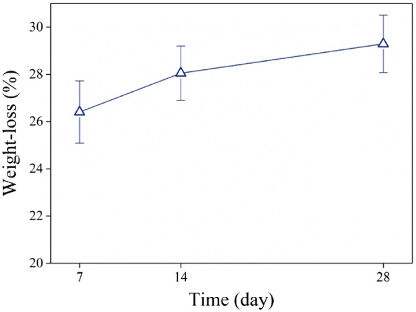
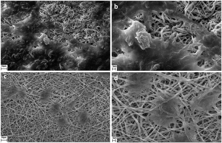
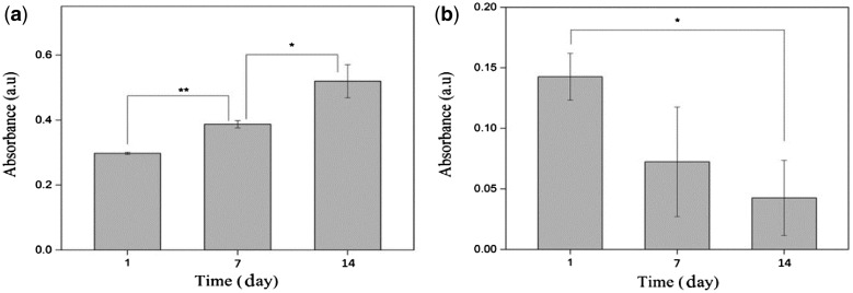
The aim of this study is to investigate the applicability of poly(lactic-co-glycolic acid) (PLGA)/collagen composite scaffold for skin tissue engineering. PLGA and collagen were dissolved in HFIP as a common solvent and fibrous scaffolds were prepared by electrospinning method. The scaffolds were characterized by scanning electron microscopy (SEM), FTIR spectroscopy, mercury porosimetry, tensile strength, biocompatibility assays and Biodegradation. Cytotoxicity and cell adhesion were tested for two cell line groups, human dermal fibroblast (HDF) and human keratinocyte (HaCat). SEM images showed appropriate cell adhesion to the scaffold for both cell lines. MTT assays indicated that the cell viability of HDF cells increased with time, but the number of HaCat cells decreased after 14 days. The ultimate tensile strength was suitable for skin substitute application, but its elongation at break was rather low. For successful clinical application of the PLGA/collagen scaffold, some properties especially mechanical strain needs to be improved.
Keywords: biocompatibility; composite scaffold; skin cells; tissue engineering.
Figures
References
-
- Powell HM, Supp DM, Boyce ST.. Influence of electrospun collagen on wound contraction of engineered skin substitutes. Biomaterials 2008;29:834–43. - PubMed
-
- Rho KS, Jeong L, Lee G. et al. Electrospinning of collagen nanofibers: effects on the behavior of normal human keratinocytes and early-stage wound healing. Biomaterials 2006;27:1452–61. - PubMed
-
- Li W-J, Laurencin CT, Caterson EJ. et al. Electrospun nanofibrous structure: a novel scaffold for tissue engineering. J Biomed Mater Res 2002;60:613–21. - PubMed
-
- Matthews JA, Wnek GE, Simpson DG. et al. Electrospinning of collagen nanofibers. Biomacromolecules 2002;3:232–8. - PubMed
-
- Chen JP, Chang GY, Chen JK.. Electrospun collagen/chitosan nanofibrous membrane as wound dressing. Colloids Surfaces A Physicochem Eng Asp 2008;313-314:183–8.
LinkOut - more resources
Full Text Sources
Other Literature Sources
