

Accurate diagnosis of prenatal cleft lip/palate by understanding the embryology
- PMID: 29026689
- PMCID: PMC5618146
- DOI: 10.5662/wjm.v7.i3.93
Accurate diagnosis of prenatal cleft lip/palate by understanding the embryology
Abstract
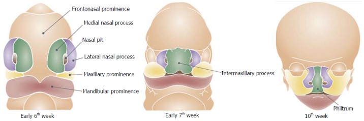
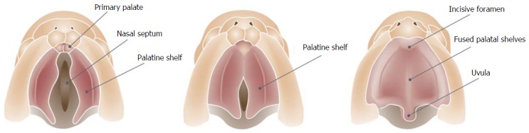
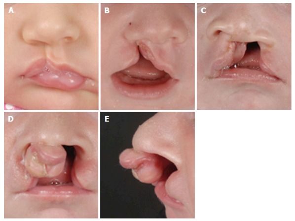
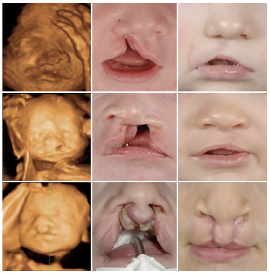
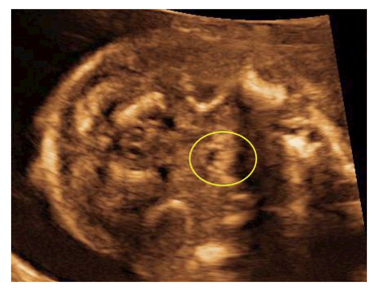
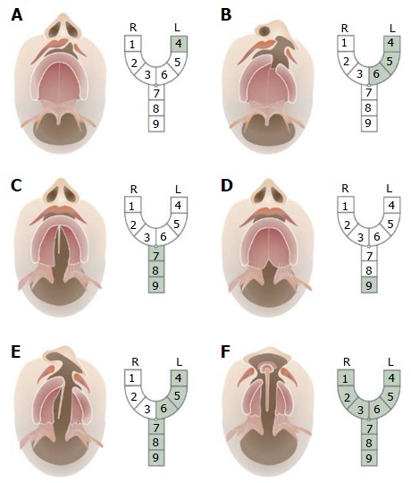
Cleft lip with or without cleft palate (CP) is one of the most common congenital malformations. Ultrasonographers involved in the routine 20-wk ultrasound screening could encounter these malformations. The face and palate develop in a very characteristic way. For ultrasonographers involved in screening these patients it is crucial to have a thorough understanding of the embryology of the face. This could help them to make a more accurate diagnosis and save time during the ultrasound. Subsequently, the current postnatal classification will be discussed to facilitate the communication with the CP teams.
Keywords: Cleft lip; Cleft palate; Embryology face; Orofacial clefts; Ultrasound.
Conflict of interest statement
Conflict-of-interest statement: The authors declare that they have no conflict of interest.
Figures
References
-
- Mulliken JB. The changing faces of children with cleft lip and palate. N Engl J Med. 2004;351:745–747. - PubMed
-
- Merritt L. Part 1. Understanding the embryology and genetics of cleft lip and palate. Adv Neonatal Care. 2005;5:64–71. - PubMed
-
- Wong FK, Hagg U. An update on the aetiology of orofacial clefts. Hong Kong Med J. 2004;10:331–336. - PubMed
-
- Böhmer AC, Mangold E, Tessmann P, Mossey PA, Steegers-Theunissen RP, Lindemans J, Bouwman-Both M, Rubini M, Franceschelli P, Aiello V, et al. Analysis of susceptibility loci for nonsyndromic orofacial clefting in a European trio sample. Am J Med Genet A. 2013;161A:2545–2549. - PubMed
Publication types
LinkOut - more resources
Full Text Sources
Other Literature Sources
Medical
Miscellaneous
