Review
. 2018:414:193-213.
doi: 10.1007/82_2017_54.

ER Protein Quality Control and the Unfolded Protein Response in the Heart

Affiliations
Review

ER Protein Quality Control and the Unfolded Protein Response in the Heart

A Arrieta et al. Curr Top Microbiol Immunol. 2018.

Abstract

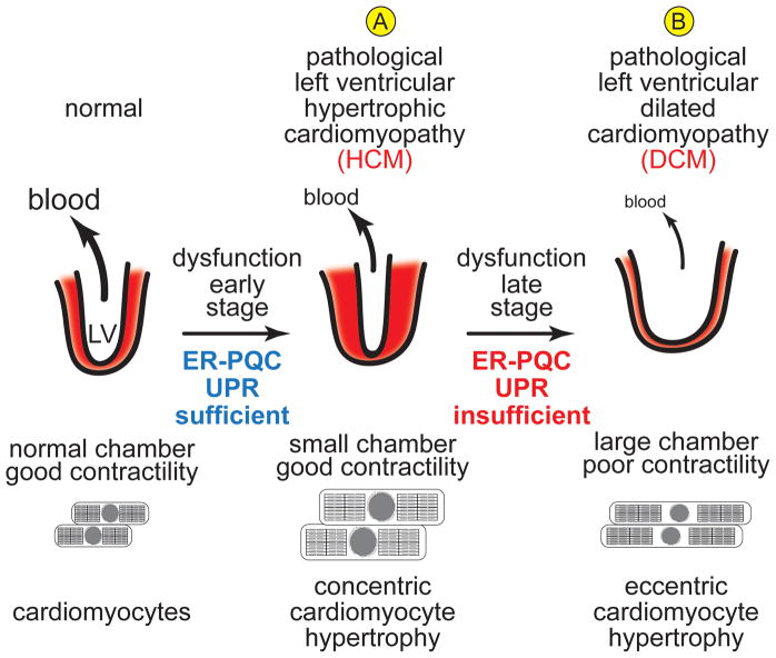
Cardiac myocytes are the cells responsible for the robust ability of the heart to pump blood throughout the circulatory system. Cardiac myocytes grow in response to a variety of physiological and pathological conditions; this growth challenges endoplasmic reticulum-protein quality control (ER-PQC), a major feature of which includes the unfolded protein response (UPR). ER-PQC and the UPR in cardiac myocytes growing under physiological conditions, including normal development, exercise, and pregnancy, are sufficient to support hypertrophic growth of each cardiac myocyte. However, the ER-PQC and UPR are insufficient to respond to the challenge of cardiac myocyte growth under pathological conditions, including myocardial infarction and heart failure. In part, this insufficiency is due to a continual decline in the expression levels of important adaptive UPR components as a function of age and during myocardial pathology. This chapter will discuss the physiological and pathological conditions unique to the heart that involves ER-PQC, and whether the UPR is adaptive or maladaptive under these circumstances.


Figures

Figure 1
Figure 1. Effects of Cardiac Pathology on ER Stress and the Unfolded Protein Response in Cardiac Myocytes
Shown is a diagram of the rough ER with attached ribosomes translating mRNAs that encode ER luminal proteins. Conditions that impair the folding of nascent ER proteins, which include ischemia, ischemia/reperfusion and hypertrophy can result in ER stress (A). Under non-stressed conditions, the ER-luminal chaperone, glucose-regulated protein 78 kDa (GRP78) associates with the luminal domains of the 3 proximal effectors of ER stress, PERK (B), IRE-1 (C) and ATF6 (D). Upon ER stress, GRP78 relocates from the luminal domains of these proteins to misfolded proteins and either facilitates their folding, or escorts them to the degradation machinery (E). The disassociation of GRP78 from PERK, IRE-1, as well as the binding of misfolded proteins to IRE-1 and, possibly to PERK, allows their oligomerization, which fosters trans-phosphorylation and activation of these effectors. In the case of ATF6, dissociation of GRP78 and the association of escorts, such as thrombospondin 4, facilitate the movement of ATF6 to the Golgi, where it is cleaved by site 1 and site 2 proteases that reside in the Golgi. The resulting N-terminal fragment is liberated from the Golgi, translocates to the nucleus, and binds to ER stress response elements in ER stress response (ERSR) genes, and regulates their transcription (D). Activated PERK (B) phosphorylates eIF2α, which fosters transient global translational repression and the translation of the ATF4 mRNA from an alternate start site to generate active ATF4 using an alternate open reading frame (ORF). While PERK appears to be cardioprotective, the ATF4-mediated induction of CHOP is pro-apoptotic in the heart (F). Activated IRE-1 splices the unspliced form of XBP1 mRNA (XBP1u mRNA) to generate a splice variant form (XBP1s mRNA) that encodes the active transcription factor, XBP1s (C). Like ATF6, XBP1s and ATF6 translocate to the nucleus, and bind to various types of regulator elements in ERSRs to regulate their expression. One important group of XBP1s genes in the heart are those that increase cytosolic protein O-GlcNAcylation, which is cardioprotective (G). Depending on whether ER stress and UPR gene induction is acute or chronic, as with other tissues, in the heart the results are either adaptive (acute), oriented toward resolution of ER stress and cell survival (G,H), or maladaptive (chronic), activating death pathways (F).
Figure 2
Figure 2. Sarco/endoplasmic Reticulum Network in a Cardiac Myocyte
Shown is a diagram of a cardiac myocyte depicting the relationships between the sarcoplasmic reticulum (SR) involved in contractile calcium handling (A), the perinuclear ER involved in secreted and membrane protein synthesis (B), the SR that may be involved in secreted and membrane protein synthesis (C), the perinuclear ER involved in nuclear calcium signaling (D) and the perinuclear ER involved in local cytosolic calcium signaling (E). Also shown are the actin and myosin myofilaments that comprise the contractile apparatus of cardiac myocytes.
Figure 3
Figure 3. Growth of the Left Ventricle during Pre- and Post-natal Development
Cross sections of the mammalian left ventricle (LV) of the heart are shown diagrammatically, with the relative size depicting the changes in LV mass at different stages of development in the newborn (A) or the adult (B). The red areas are representations of the myocardium. The relative sizes of the arrows and blood represent the relative amounts of blood pumped, or ejected by the LV at different stages of development. The cardiac myocyte diagrams below each LV represent the relative changes in number and size during development. Since increases in LV muscle mass by hyperplasic or hypertrophic growth require increases in protein-folding machinery, and since there is not ER stress or cell death associated with LV myocyte growth during pre- and post-natal development, endoplasmic reticulum-protein quality control (ER-PQC) and unfolded protein response (UPR) machinery are sufficient to support myocardial growth.
Figure 4
Figure 4. Growth of the Left Ventricle during Exercise and Pregnancy
Cross sections of the LV and cardiomyocytes are depicted, as in Figure 3. During exercise and pregnancy the LV grows adaptively by concentric hypertrophy of each cardiomyocyte, i.e. increased diameter but not increased length, while the number of myocytes remains unchanged. Since this growth is adaptive, and is not associated with ER stress or myocyte death, the ER-PQC and UPR are sufficient to support the growth.
Figure 5
Figure 5. Effect of Ischemia, Myocardia infarction, Mutations, Drugs and Pathogens on Cardiac Contractility
(A) Myocardial infarction (MI) results in irreparable damage to the myocardium, which decreases LV contractility. Ischemic cardiomyocytes that eventually die from ischemia have insufficient ER-PQC and UPR machinery. (B) Mutations, drugs, mostly anti-neoplastic agents, and pathogens, such as viruses, also impair cardiomyocyte contractility. These insults are associated with an insufficient ER-PQC and UPR, leading to maladaptive ER stress, which contributes to myocyte dropout by apoptosis.
Figure 6
Figure 6. Development of Cardiomyopathy in the Diseased Heart
Ischemia, MI, mutations, drugs and pathogens often lead to (A) hypertrophic cardiomyopathy (HCM), a condition in which each LV myocyte grows concentrically, i.e. increased diameter. This hypertrophic growth of the LV does not lead to increased pump function, and in many cases LV pump function is decreased, as the LV wall growth is so extensive that the chamber size is decreased. This hypertrophy is considered pathological because it usually leads to further remodeling and deterioration of LV structure and LV dysfunction in the late stage of pathology, associated with cardiomyocyte apoptosis, as well as an eccentric growth of remaining cardiomyocyte, i.e. increased length, which does not enhance cardiomyocyte contractility. This results in a dilation of the LV so chamber size increases, but since LV muscle mass is decreased, the ability of the LV to pump blood is severely impaired, leading to dilated hypertrophic cardiomyopathy (DCM) and eventually to heart failure (B). In the early stage of the disease, the ER-PQC and UPR are sufficient to handle cardiomyocyte growth; however, in the late stage of the disease, maladaptive ER stress ensues, and the ER-PQC and UPR are not sufficient to maintain LV structure and function.

References

    1. Abarbanell AM, Herrmann JL, Weil BR, Wang Y, Tan J, Moberly SP, Fiege JW, Meldrum DR. Animal models of myocardial and vascular injury. J Surg Res. 2010;162(2):239–249. doi: 10.1016/j.jss.2009.06.021. - DOI - PubMed
    1. Banerjee I, Fuseler JW, Price RL, Borg TK, Baudino TA. Determination of cell types and numbers during cardiac development in the neonatal and adult rat and mouse. Am J Physiol Heart Circ Physiol. 2007;293(3):H1883–1891. doi: 10.1152/ajpheart.00514.2007. - DOI - PubMed
    1. Belmont PJ, Tadimalla A, Chen WJ, Martindale JJ, Thuerauf DJ, Marcinko M, Gude N, Sussman MA, Glembotski CC. Coordination of growth and endoplasmic reticulum stress signaling by regulator of calcineurin 1 (RCAN1), a novel ATF6-inducible gene. J Biol Chem. 2008;283(20):14012–14021. doi: 10.1074/jbc.M709776200. M709776200 [pii] - DOI - PMC - PubMed
    1. Bers DM. Cardiac excitation-contraction coupling. Nature. 2002;415(6868):198–205. doi: 10.1038/415198a. - DOI - PubMed
    1. Bers DM. Calcium cycling and signaling in cardiac myocytes. Annu Rev Physiol. 2008;70:23–49. doi: 10.1146/annurev.physiol.70.113006.100455. - DOI - PubMed

MeSH terms

LinkOut - more resources