Review
. 2017 Nov;280(1):149-164.
doi: 10.1111/imr.12587.

Efferocytosis of dying cells differentially modulate immunological outcomes in tumor microenvironment

Affiliations
Review

Efferocytosis of dying cells differentially modulate immunological outcomes in tumor microenvironment

Sushil Kumar et al. Immunol Rev. 2017 Nov.

Abstract

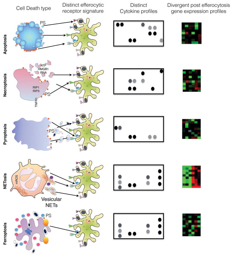
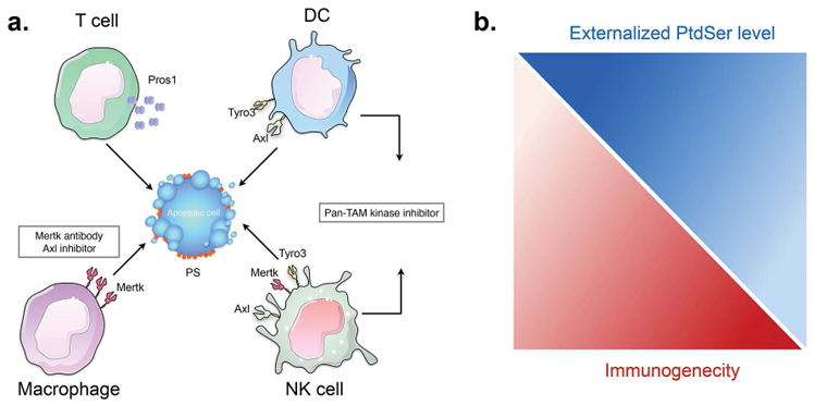
Programmed cell death (apoptosis) is an integral part of tissue homeostasis in complex organisms, allowing for tissue turnover, repair, and renewal while simultaneously inhibiting the release of self antigens and danger signals from apoptotic cell-derived constituents that can result in immune activation, inflammation, and autoimmunity. Unlike cells in culture, the physiological fate of cells that die by apoptosis in vivo is their rapid recognition and engulfment by phagocytic cells (a process called efferocytosis). To this end, apoptotic cells express specific eat-me signals, such as externalized phosphatidylserine (PS), that are recognized in a specific context by receptors to initiate signaling pathways for engulfment. The importance of carefully regulated recognition and clearance pathways is evident in the spectrum of inflammatory and autoimmune disorders caused by defects in PS receptors and signaling molecules. However, in recent years, several additional cell death pathways have emerged, including immunogenic cell death, necroptosis, pyroptosis, and netosis that interweave different cell death pathways with distinct innate and adaptive responses from classical apoptosis that can shape long-term host immunity. In this review, we discuss the role of different cell death pathways in terms of their immune potential outcomes specifically resulting in specific cell corpse/phagocyte interactions (phagocytic synapses) that impinge on host immunity, with a main emphasis on tolerance and cancer immunotherapy.

Keywords: cell death; immunogenic cell death; phosphatidylserine; tolerance; tumor immunity.


Conflict of interest statement

Disclosure of potential conflicts of interest:

No potential conflicts of interest were disclosed by the authors.

Figures

Figure 1
Figure 1. Genetic regulation of cell death and engulfment pathways in C.elegans
During apoptotic cell (left panel), caspase (CED-3) activation leads to a cell death cascade that includes pathways leading to CED7 and CED8-mediated externalization of PS. PS externalization on dying cells is recognized by a series of PS receptors and PS bridging molecules that detect the dying cells, that in turn engage signaling pathways involving CED-2, CED-5 and CED12 that lead to CED-10 activation, for subsequent engulfment and degradation of the apoptotic cell (right panel).
Figure 2
Figure 2. Detection of PS via PS receptors, and signaling via Crk, DOCK180, Rac1 in phagocytic cells
Apoptotic cells express “eat-me” signals, such as externalized PS in response to apoptotic stimuli. Externalized PS, in turn, is recognized directly by a variety of PS receptors expressed on phagocytes and bridging molecules such C1q, MFG-E8, Gas6, and Pros1. Many PS receptors and PS bridging molecules, when engaged bv apoptotic cells, subsequently lead to activation of an evolutionarily conserved Crk-DOCK-Rac1 pathway.
Figure 3
Figure 3. Model for the differential engulfment outcomes resulting from PS externalization via caspase activation (Xkr8/ATP11C) and via cellular stress (TMEM16F)
As developed in the text, PS is externalized via the activity of distinct classes of lipid transporters during apoptosis versus cellular stress. During apoptosis, PS becomes externalized concomitant with caspase activation by the inactivation of ATP11C (flippase), and the concomitant activation of Xkr8 (scramblase), leading to irreversible PS externalization. By contrast, during cellular stress, activation of calcium-dependent TMEM16F (scramblase) leads to transient and reversible externalization of PS. In addition to the issue of reversibility of PS externalization, other factors, such as the down-regulation of the don’t-eat me receptor CD47, and the lateral movement and clustering of PS, likely contribute differential signaling of externalized PS have been implicated in the differential fates of externalized PS (see ref 59, 69).
Figure 4
Figure 4. PS receptors (Tyro3, Axl, and Mertk) differentially impact the tumor microenvironment via the interaction of apoptotic cells
(A) The expression of TAMs and their ligands (Gas6 and Pros1) on infiltrating tumor-associated immune subsets may act as immune checkpoint inhibitors that promote tolerogenic signals in the tumor microenvironment. Examples include; Mertk expressed on macrophages, Tyro3 and Axl expressed on DCs, and Tyro3, Axl, and Mertk expressed on NK cells, and Pros1 expressed on activated T cells. In recent years, the development of TAM antagonists are being assessed as myeloid checkpoint inhibitors in cancer. In some scenario’s, inhibition of Mertk on macrophages might be expected to skew efferocytosis towards antigen presenting cells (DCs) that establish tumor immunity. In other cases, pan TAM inhibitors might stimulate global immunogenic outcomes by blocking multiple inhibitory signals. (B). Externalized PS is a double-edge sword in the tumor microenvironment. In this model, tumors with high deposition of externalized PS will have low immunogenicity, and vice versa, such that PS targeting antibodies are expected to have important therapeutic value in cancer immunotherapy
Figure 5
Figure 5. Decoding cell death modules by phagocytic cells into efferocytic synapses
Cells that have undergone cell death through various mechanisms include apoptosis, necroptosis, pyroptosis, NETosis, and ferroptosis. These different death modalities produce distinct proteomic and lipidomic surface components that in turn engage specific repertoires of surface receptors on the engulfing cells, akin to specific phagocytic synapses. Intuitively, these ligand receptor interactions will produce signature gene expression and cytokine profiles. Such systems biology platforms may help better define different cell death platforms and how they influence the tumor microenvironment.

References

    1. Elliott MR, Ravichandran KS. Clearance of apoptotic cells: implications in health and disease. J Cell Biol. 2010;189:1059–1070. - PMC - PubMed
    1. Savill J, Dransfield I, Gregory C, Haslett C. A blast from the past: clearance of apoptotic cells regulates immune responses. Nat Rev Immunol. 2002;2:965–975. - PubMed
    1. Savill J, Fadok V. Corpse clearance defines the meaning of cell death. Nature. 2000;407:784–788. - PubMed
    1. Voll RE, Herrmann M, Roth EA, et al. Immunosuppressive effects of apoptotic cells. Nature. 1997;390:350–351. - PubMed
    1. Duvall E, Wyllie AH, Morris RG. Macrophage recognition of cells undergoing programmed cell death (apoptosis) Immunology. 1985;56:351–358. - PMC - PubMed

Publication types

Substances

LinkOut - more resources