. 2018 Jan;39(1):124-139.
doi: 10.1002/humu.23354. Epub 2017 Nov 2.

Functional characterization of novel NR5A1 variants reveals multiple complex roles in disorders of sex development

Affiliations

Functional characterization of novel NR5A1 variants reveals multiple complex roles in disorders of sex development

Gorjana Robevska et al. Hum Mutat. 2018 Jan.

Abstract

Variants in the NR5A1 gene encoding SF1 have been described in a diverse spectrum of disorders of sex development (DSD). Recently, we reported the use of a targeted gene panel for DSD where we identified 15 individuals with a variant in NR5A1, nine of which are novel. Here, we examine the functional effect of these changes in relation to the patient phenotype. All novel variants tested had reduced trans-activational activity, while several had altered protein level, localization, or conformation. In addition, we found evidence of new roles for SF1 protein domains including a region within the ligand binding domain that appears to contribute to SF1 regulation of Müllerian development. There was little correlation between the severity of the phenotype and the nature of the NR5A1 variant. We report two familial cases of NR5A1 deficiency with evidence of variable expressivity; we also report on individuals with oligogenic inheritance. Finally, we found that the nature of the NR5A1 variant does not inform patient outcomes (including pubertal androgenization and malignancy risk). This study adds nine novel pathogenic NR5A1 variants to the pool of diagnostic variants. It highlights a greater need for understanding the complexity of SF1 function and the additional factors that contribute.

Keywords: NR5A1; disorders of sex development; genotype-phenotype correlation; mutation; oligogenic; variable expressivity.


Figures

Figure 1
Figure 1
Variants in SF1 identified in patients with disorders of sex development using massively parallel sequencing. A: A schematic representation of the predicted protein structure of SF1 showing the approximate location of the variants identified in a cohort of DSD patients. The protein domains are as follows: DNA binding domain (DBD) containing two zinc finger motifs (Zn) and the Fushi‐tarazu factor 1 box (Ftz‐F1), the hinge region and ligand binding domain (LBD). P Box, T‐box, A‐box, as well as two activational domains—AF1, AF2. Patient variants (with patient number denoted by P#) are shown. Five missense mutations were identified in our cohort of 46,XY DSD patients, some of which were recurrent. Two fell within the DBD (p.G35D, p.R84H) one in the hinge region (p.G178R), two were in the LBD (p.H310D and p.D364Y). Two in‐frame deletions were found, one within the second zinc finger motif (p.47_54del) and the other in the LBD (p.K372del); two frame‐shifts were identified (p.R89Gfs*17 and p.L209Cfs*87) as well as a nonsense mutation at position 211. Blue boxes denote single nucleotide variants, yellow are in‐frame deletions and pink are variants that cause a frame‐shift and the black box is a nonsense mutation. B: Evolutionary conservation of the SF1 protein (the last 40 amino acids are not shown). All variants are indicated, in‐frame deletions are shaded in yellow while frame shifts are indicated with pink shading at the point of the new transcript, in addition the zinc finger motifs, nuclear localization signal (NLS), and the conserved helices which are in the LBD are shown
Figure 2
Figure 2
Splice site mutations identified in 46,XY individuals. A: A schematic representation of NR5A1, showing the six coding exons and the position of the two splice site acceptor mutations identified. B and C:Human Splicing Finder (Desmet et al., 2009) and SpliceAid 2 (Piva et al., 2012) were used to analyze the consequences of the two splice site acceptor mutations. Both of which suggest that wild‐type splicing would be affected
Figure 3
Figure 3
Reduced trans‐activational activity of SF1 variants. The trans‐activational activity of each variant compared with wild‐type SF1 was tested using a dual‐luciferase reporter assay in COS‐7 cells. Each SF1 variant was transfected either alone (blue bars), with hSRY (red bars) or with hSOX9 (green bars) and transcriptional activity was measured by activation of the mTesco promoter driven luciferase reporter (p.GL4). Empty vector, in place of SF1 was run in each condition as a negative control. Briefly wild‐type SF1 alone shows only low activation of mTesco, however transactivation activity increased around four times with the addition of SOX9. All SF1 variants tested show a significant decrease in trans‐activational activity with SOX9 and with SRY. Complete loss of trans‐activational activity was noted for the proteins with missense mutations located in the LBD (p.H310D and p.D364Y), the p.47_54 in‐frame deletion, as well as both frame‐shift mutations assayed (p.R89Gfs*17 and p.L209Cfs*87). The nonsense mutation SF1 (p.[P210Q;Y211*]) seemed to retain a low level of activity. Data represent the mean with standard error of four independent experiments performed in duplicate transfections. Unpaired t‐test was applied and for **P value < 0.005; *P value < 0.05
Figure 4
Figure 4
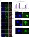
A: Protein expression is affected in some SF1 variants. Protein expression of each variant and wild‐type SF1 was assessed in COS‐7 cells with an SF1 antibody (green). Cells were transfected with an equal amount of SF1 expression vector (wild‐type or variant). Nuclear counterstaining was performed with DAPI (blue) and the cytoskeleton was stained with actin (red). Wild‐type SF1 showed strong nuclear staining with nucleolar exclusions. All SF1 expression vectors with missense mutations and in‐frame deletion were found to be expressed while none of the truncated proteins were detected (p.R89Gfs*17, p.L209Cfs*87, and p.[P210Q;Y211*]). B: This was quantified as the number of SF1 expressing nuclei per image (2–10×, two cropped areas c.f. 40× displayed in the image). C: Mutant protein p.G35D was dispersed through the cytosol; while the in‐frame deletion affecting the Zn II motif was clumped in sub‐nuclear aggregates; and p.R84H seemed to be concentrated on the nuclear border
Figure 5
Figure 5
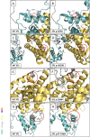
Conformational changes in SF1 variant proteins. To investigate the potential impact of each variant on protein conformation, we performed an in silico prediction with the WT SF1 and each variant using I‐Tasser and PyMol modeling software. For variants identified in patients with CGD or PGD, the side chains are shown on the implicated residues. A: Wild‐type Gly at position 35 falls within a highly specific 34KGFFK38 motif (motif side chains shown), where both lysine residues are thought to be subjected to post translational acetylation. The glycine residue is substitute with a larger, negatively charged aspartic acid, (B) decreasing the distance between the two lysine residues at position 35 and 38, from 10.4 to 7.1 A. C: Wild‐type His residue at position 310 falls within the highly conserved alpha helix 5 of LBD (circled) and is a large, neutral amino acid commonly involved in stacking, (D) while mutant Asp is negatively charged and much smaller. E: The Asp to Tyr transition at position 364, which falls within the LBD, adjacent to a highly conserved alpha helix 8. The wild‐type Asp is quite small and secondary structure is predicted be at a turn, (F) unlike the bulky Tyr, which would not have that secondary structure. G: Eight amino acid deletion in the second Zn finger motif of the DNA binding domain, (H) results in a loss of one of the cysteine residues, which is crucial for the Zn2+ interaction and possible loss of stability and ability to bind conical 6‐bp HRE

References

    1. Achermann, J. C. , Ito, M. , Ito, M. , Hindmarsh, P. , & Jameson, J. L. 1999. A mutation in the gene encoding steroidogenic factor‐1 causes XY sex reversal and adrenal failure in humans. Nature Genetics, 22, 125–126. - PubMed
    1. Allali, S. , Muller, J‐B. , Brauner, R. , Lourenço, D. , Boudjenah, R. , Karageorgou, V. , … Bashamboo, A. (2011). Mutation analysis of NR5A1 encoding steroidogenic factor 1 in 77 patients with 46, XY disorders of sex development (DSD) including hypospadias. PLoS One, 6, e24117–e2418. - PMC - PubMed
    1. Arango, N. A. , Lovell‐Badge, R. , & Behringer, R. R. (1999). Targeted mutagenesis of the endogenous mouse. Cell, 99, 409–419. - PubMed
    1. Baetens, D. , Mladenov, W. , Delle Chiaie, B. , Menten, B. , Desloovere, A. , Iotova, V. , … Cools, M. (2014). Extensive clinical, hormonal and genetic screening in a large consecutive series of 46,XY neonates and infants with atypical sexual development. Orphanet Journal of Rare Diseases, 9, 209. - PMC - PubMed
    1. Barbaro, M. , Cools, M. , Looijenga, L. H. J. , Drop, S. L. S. , & Wedell, A. (2011). Partial deletion of the NR5A1 (SF1) gene detected by synthetic probe MLPA in a patient with XY gonadal disorder of sex development. Sexual Development, 5, 181–187. - PubMed

MeSH terms

LinkOut - more resources