. 2017 Nov 9;60(21):8982-8988.
doi: 10.1021/acs.jmedchem.7b01221. Epub 2017 Oct 27.

Structure-Based Design of Non-natural Macrocyclic Peptides That Inhibit Protein-Protein Interactions

Affiliations

Structure-Based Design of Non-natural Macrocyclic Peptides That Inhibit Protein-Protein Interactions

Dennis M Krüger et al. J Med Chem. .

Abstract

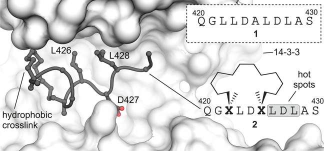
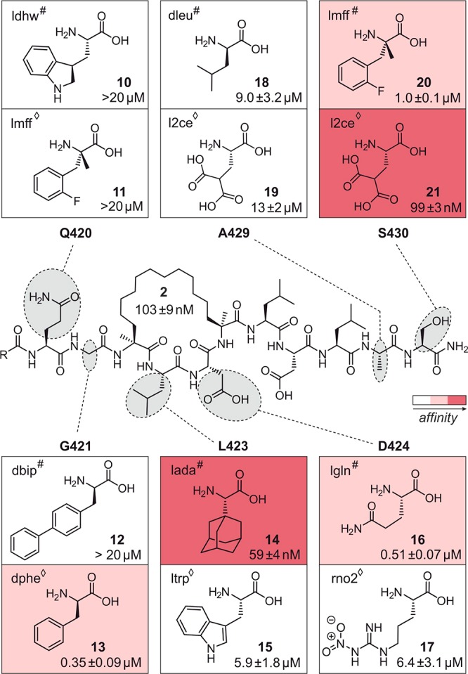
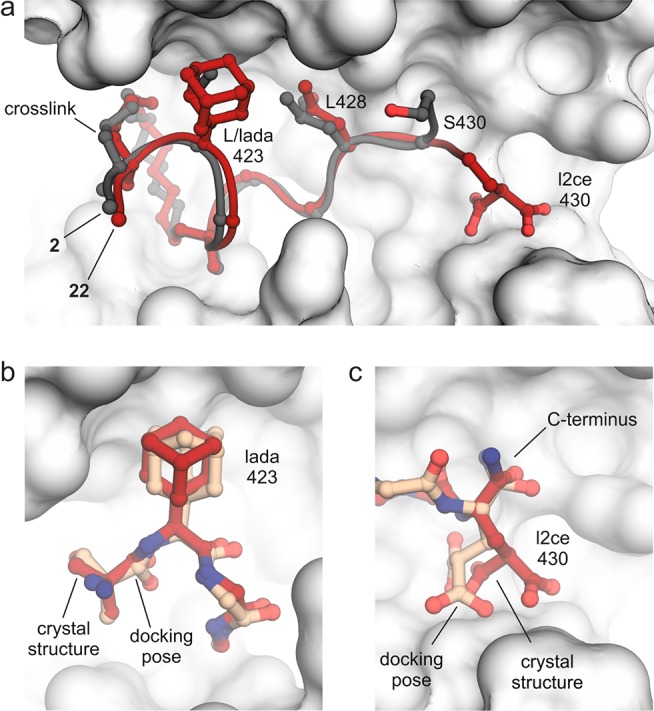
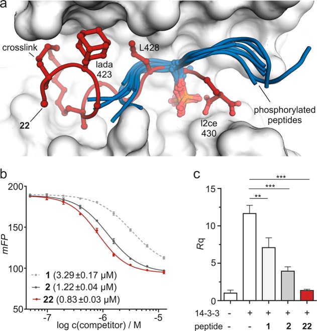
Macrocyclic peptides can interfere with challenging biomolecular targets including protein-protein interactions. Whereas there are various approaches that facilitate the identification of peptide-derived ligands, their evolution into higher affinity binders remains a major hurdle. We report a virtual screen based on molecular docking that allows the affinity maturation of macrocyclic peptides taking non-natural amino acids into consideration. These macrocycles bear large and flexible substituents that usually complicate the use of docking approaches. A virtual library containing more than 1400 structures was screened against the target focusing on docking poses with the core structure resembling a known bioactive conformation. Based on this screen, a macrocyclic peptide 22 involving two non-natural amino acids was evolved showing increased target affinity and biological activity. Predicted binding modes were verified by X-ray crystallography. The presented workflow allows the screening of large macrocyclic peptides with diverse modifications thereby expanding the accessible chemical space and reducing synthetic efforts.


Conflict of interest statement

The authors declare no competing financial interest.

Figures

Figure 1
Figure 1
Sequence of linear peptide 1 and crystal structure of cyclic peptide 2 (dark gray) bound to 14–3–3ζ (light gray, PDB ID 4n84). Cross-link and hotspot residues (L426, D427, and L428) are shown explicitly. Peptide sequence of 2 and chemical structure of cross-link are shown (Residues are numbered in accordance to PDB ID 4n84).
Figure 2
Figure 2
Chemical structure of 2 with varied residues highlighted in gray. Selected 12 variations are shown (two per position: one obtained from ChemScore (#) and the other one from ASP (◊) hit list). Experimentally determined dissociation constants (Kd) for corresponding peptides 1021 with 14–3–3ζ are given (for details see Figure S7). Variations are color coded in accordance to their affinity for 14–3–3ζ (Kd rages: dark red, <0.1 μM; light red, 0.1–1 μM; white, >1 μM).
Figure 3
Figure 3
(a) Overlay of crystal structures of 2 (gray, PDB ID 4n84) and 22 (red, PDB ID 5jm4) when bound to 14–3–3ζ (light gray surface representation). Peptide backbones are shown as ribbons. Varied side chains (423 and 430), hotspot residue L428, and cross-link are shown explicitly. (b,c) Superimposition of 22 crystal structure (red, PDB ID 5jm4) with predicted structure (orange, pose with highest score) of lada- and l2ce-modified peptide, respectively. Amino acid of interest and backbone of neighboring amino acids are shown explicitly.
Figure 4
Figure 4
(a) Superimposition of 22 (red, PDB ID 5jm4) and selected phosphorylated peptides (blue, PDB IDs iqja, 1ywt, 2bn5, 2btp, 2c74, 2npm, 2v7d, 3e6y, and 3nkx) bound to 14–3–3. Peptide backbones are shown as ribbons and important side chains explicitly. (b) FP competition using labeled Raf-peptide as tracer (10 nM with 2 μM 14–3–3) and nonlabeled versions of 1, 2, and 22 (including their IC50 values). (c) Inhibition of MMP1 transcription in a cell-based assay after pathway activation with 14–3–3ζ (c = 200 nM). Cells were treated for 24 h in the absence and presence of 1, 2, or 22 (20 μM). Expression levels of mRNA were measured by quantitative real time PCR (for details, see Supporting Information).

References

    1. Villar E. A.; Beglov D.; Chennamadhavuni S.; Porco J. A. Jr.; Kozakov D.; Vajda S.; Whitty A. How proteins bind macrocycles. Nat. Chem. Biol. 2014, 10 (9), 723–731. 10.1038/nchembio.1584. - DOI - PMC - PubMed
    1. Pelay-Gimeno M.; Glas A.; Koch O.; Grossmann T. N. Structure-based design of inhibitors of protein-protein interactions: mimicking peptide binding epitopes. Angew. Chem., Int. Ed. 2015, 54 (31), 8896–8927. 10.1002/anie.201412070. - DOI - PMC - PubMed
    1. Sperandio O.; Reynes C. H.; Camproux A.-C.; Villoutreix B. O. Rationalizing the chemical space of protein-protein interaction inhibitors. Drug Discovery Today 2010, 15 (5–6), 220–229. 10.1016/j.drudis.2009.11.007. - DOI - PubMed
    1. Nevola L.; Giralt E. Modulating protein-protein interactions: the potential of peptides. Chem. Commun. 2015, 51 (16), 3302–3315. 10.1039/C4CC08565E. - DOI - PubMed
    1. Milroy L. G.; Grossmann T. N.; Hennig S.; Brunsveld L.; Ottmann C. Modulators of protein-protein interactions. Chem. Rev. 2014, 114 (9), 4695–4748. 10.1021/cr400698c. - DOI - PubMed

Publication types