. 2018 Nov 1;28(11):3939-3950.
doi: 10.1093/cercor/bhx257.

Serotonin 2A Receptor Signaling Underlies LSD-induced Alteration of the Neural Response to Dynamic Changes in Music

Affiliations

Serotonin 2A Receptor Signaling Underlies LSD-induced Alteration of the Neural Response to Dynamic Changes in Music

Frederick S Barrett et al. Cereb Cortex. .

Abstract

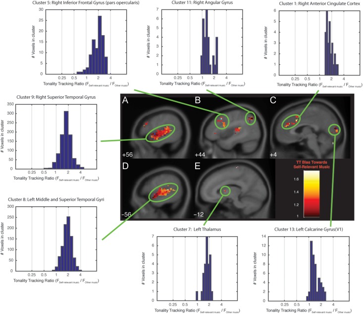
Classic psychedelic drugs (serotonin 2A, or 5HT2A, receptor agonists) have notable effects on music listening. In the current report, blood oxygen level-dependent (BOLD) signal was collected during music listening in 25 healthy adults after administration of placebo, lysergic acid diethylamide (LSD), and LSD pretreated with the 5HT2A antagonist ketanserin, to investigate the role of 5HT2A receptor signaling in the neural response to the time-varying tonal structure of music. Tonality-tracking analysis of BOLD data revealed that 5HT2A receptor signaling alters the neural response to music in brain regions supporting basic and higher-level musical and auditory processing, and areas involved in memory, emotion, and self-referential processing. This suggests a critical role of 5HT2A receptor signaling in supporting the neural tracking of dynamic tonal structure in music, as well as in supporting the associated increases in emotionality, connectedness, and meaningfulness in response to music that are commonly observed after the administration of LSD and other psychedelics. Together, these findings inform the neuropsychopharmacology of music perception and cognition, meaningful music listening experiences, and altered perception of music during psychedelic experiences.


Figures

Figure 1.
Figure 1.
Brain areas tracking the tonal structure of personally meaningful and other musical selections. Each labeled panel (AE) depicts a sagittal brain slice containing a TT cluster. Shading within each cluster indicates the TT bias for the voxels in the cluster. TT bias is the ratio of variance explained by TT regressors for personally meaningful stimuli to variance explained by the TT regressors for other stimuli (corrected for the number of stimuli in each category). A TT bias value of 1 indicates voxels that tracked personally meaningful and other stimuli equally well; greater than 1 indicates voxels that tracked personally meaningful stimuli more strongly than other stimuli. TT clusters are circled in green, and the distribution of TT bias for all voxels in that cluster is indicated in the associated histogram. Each histogram refers to a cluster listed in Supplementary Table 1. The number in the lower-left-hand corner of each sagittal slice indicates the Montreal Neurologic Institute template coordinate (in mm) of that slice in the x dimension.
Figure 2.
Figure 2.
Brain areas tracking the tonal structure of music during placebo compared to LSD. Panels and histograms as in Figure 1. Each labeled panel (AE) depicts a sagittal brain slice containing a TT cluster. The number in the lower-left-hand corner of each sagittal slice indicates the Montreal Neurologic Institute template coordinate (in mm) of that slice in the x dimension. Shading on the warm gradient within each cluster indicates voxels that are biased toward TT during placebo, and shading on the cool gradient indicates voxels that are biased toward TT during LSD. A TT bias value of 1 indicates voxels that tracked stimuli equally well during placebo and LSD; greater than 1 (plotted on the warm gradient) indicates voxels that tracked stimuli more strongly during placebo than during LSD; less than 1 (plotted on the cool gradient) indicates voxels that tracked stimuli more strongly during LSD than during placebo. Each histogram refers to a cluster listed in Supplementary Table 2.
Figure 3.
Figure 3.
Brain areas tracking the tonal structure of music during LSD compared to Ket + LSD. Panels and histograms as in Figure 1. Each labeled panel (AE) depicts a sagittal brain slice containing a TT cluster. The number in the lower-left-hand corner of each sagittal slice indicates the Montreal Neurologic Institute template coordinate (in mm) of that slice in the x dimension. Shading on the cool gradient within each cluster indicates voxels that are biased toward TT during LSD, and shading on the green gradient indicates voxels that are biased toward TT during Ket + LSD. A TT bias value of 1 indicates voxels that tracked stimuli equally well during LSD and Ket + LSD; greater than 1 (plotted on the cool gradient) indicates voxels that tracked stimuli more strongly during LSD than during Ket + LSD; less than 1 (plotted on the green gradient) indicates voxels that tracked stimuli more strongly during Ket + LSD than during LSD. Each histogram refers to a cluster listed in Supplementary Table 3.
Figure 4.
Figure 4.
Brain areas tracking the tonal structure of music during placebo compared with Ket + LSD. Panels and histograms as in Figure 1. Each labeled panel (AE) depicts a sagittal brain slice containing a TT cluster. The number in the lower-left-hand corner of each sagittal slice indicates the Montreal Neurologic Institute template coordinate (in mm) of that slice in the x dimension. Shading on the warm gradient within each cluster indicates voxels that are biased toward TT during placebo, and shading on the green gradient indicates voxels that are biased toward TT during Ket + LSD. A value of 1 indicates voxels that tracked stimuli equally well during placebo and Ket + LSD; greater than 1 (plotted on the warm gradient) indicates voxels that tracked stimuli more strongly during placebo than during Ket + LSD; less than 1 (plotted on the green gradient) indicates voxels that tracked stimuli more strongly during Ket + LSD than during placebo. Each histogram refers to a cluster listed in Supplementary Table 4.

References

    1. Amunts K, Kedo O, Kindler M, Pieperhoff P, Mohlberg H, Shah NJ, Habel U, Schneider F, Zilles K. 2005. Cytoarchitectonic mapping of the human amygdala, hippocampal region and entorhinal cortex: Intersubject variability and probability maps. Anat Embryol (Berl). 210(5–6):343–352. - PubMed
    1. Andree B, Nyberg S, Ito H, Ginovart N, Brunner F, Jaquet F, Halldin C, Farde L. 1998. Positron emission tomographic analysis of dose-dependent MDL 100,907 binding to 5-hydroxytryptamine-2A receptors in the human brain. J Clin Psychopharmacol. 18(4):317–323. - PubMed
    1. Ardila A, Bernal B, Rosselli M. 2016. How localized are language brain areas? A review of brodmann areas involvement in oral language. Arch Clin Neuropsychol. 31(1):112–122. - PubMed
    1. Barrett FS, Janata P. 2016. Neural responses to nostalgia-evoking music modeled by elements of dynamic musical structure and individual differences in affective traits. Neuropsychologia. 91:234–246. - PubMed
    1. Barrett FS, Robbins H, Smooke D, Brown JL, Griffiths RR. 2017. Qualitative and quantitative features of music reported to support peak mystical experiences during psychedelic therapy sessions. Frontiers in Psychol. 8:1238. - PMC - PubMed

Publication types

MeSH terms