. 2017 Dec 1;158(12):4317-4330.
doi: 10.1210/en.2017-00618.

Thyroid Hormone Metabolism Defects in a Mouse Model of SBP2 Deficiency

Affiliations

Thyroid Hormone Metabolism Defects in a Mouse Model of SBP2 Deficiency

Jiao Fu et al. Endocrinology. .

Abstract

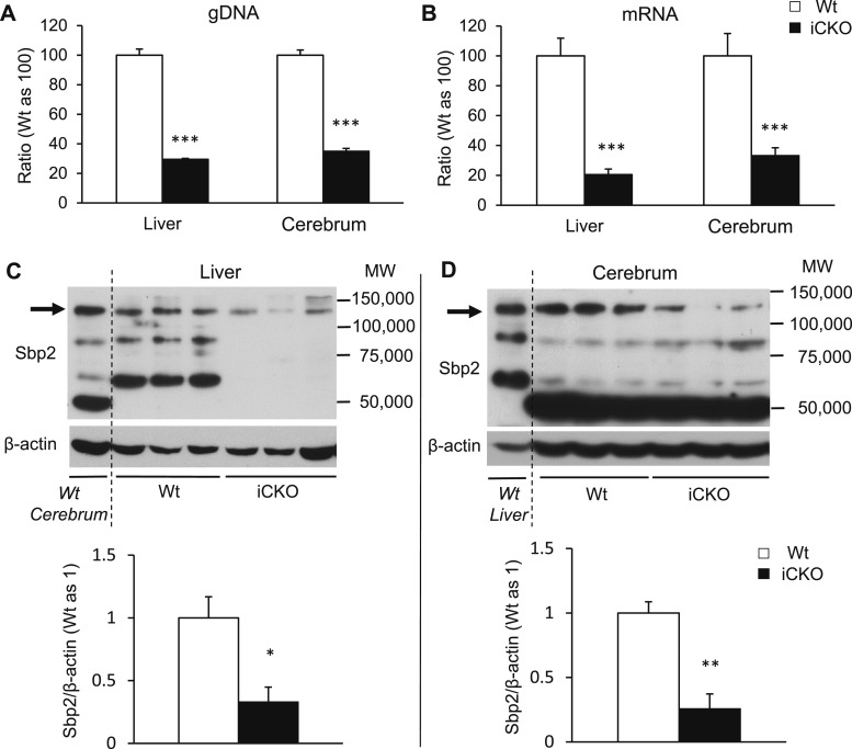
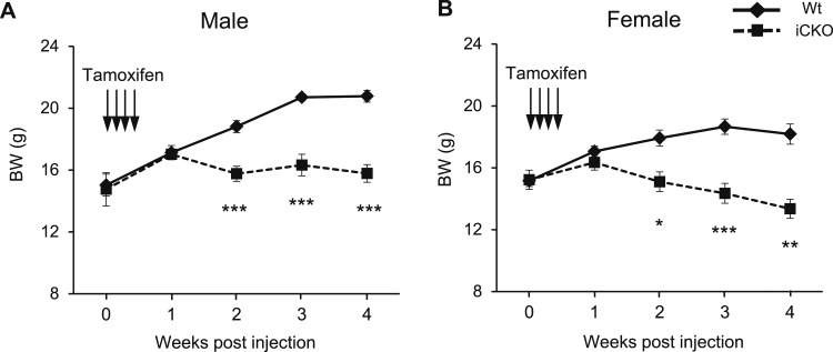
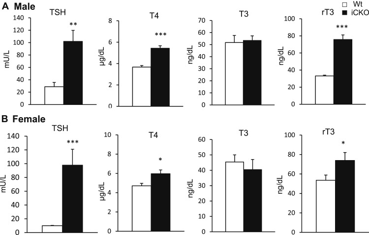
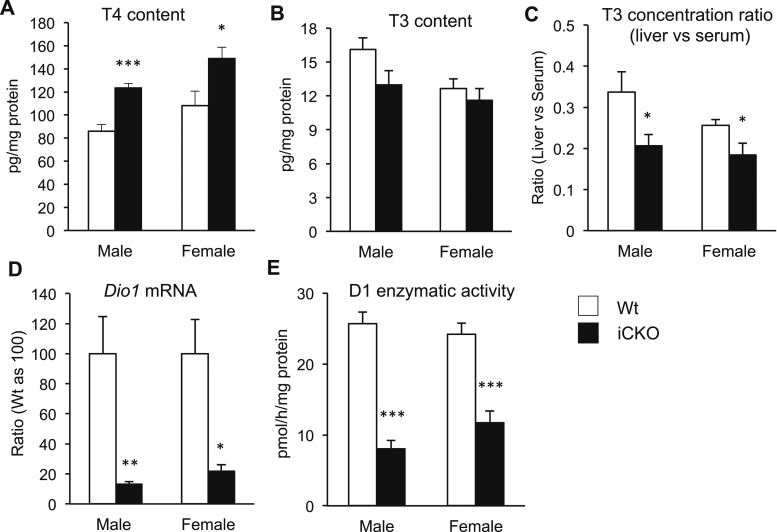
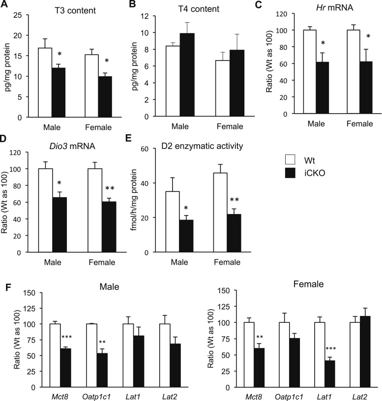
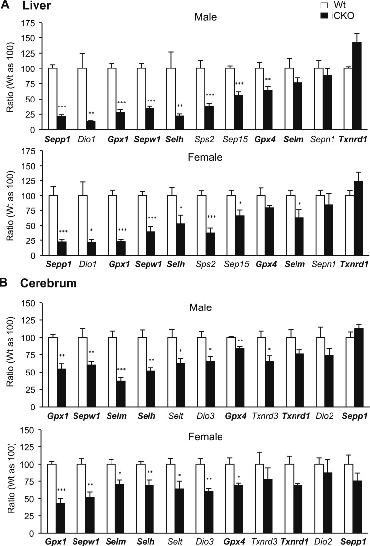
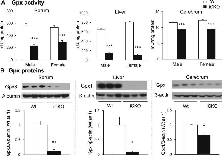
Selenocysteine insertion sequence binding protein 2 (SBP2) is an essential factor in selenoprotein synthesis. Patients with SBP2 defects have a characteristic thyroid phenotype and additional manifestations such as growth delay, male infertility, impaired motor coordination, and developmental delay. The thyroid phenotype has become pathognomonic for this defect, and putative deficiencies in the iodothyronine deiodinases selenoenzymes have been implicated. To investigate the role of SBP2 and selenoproteins in thyroid physiology and answer questions raised by the human syndrome, we generated a tamoxifen-inducible Sbp2 conditional knockout (iCKO) mouse model. These Sbp2-deficient mice have high serum thyroxine (T4), thyrotropin, and reverse triiodothyronine (T3), similar to the human phenotype of SBP2 deficiency, whereas serum T3 is normal. Their liver T4 and T3 content reflect the serum levels, and deiodinase 1 expression and enzymatic activity were decreased. In contrast, brain T3 content is decreased, indicative of local hypothyroidism, confirmed by the decreased expression of the thyroid hormone (TH) positively regulated gene hairless. Interestingly, the cerebrum T4 content did not parallel the high serum T4 levels, and the expression of TH transporters was decreased. Deiodinase 2 enzymatic activity and deiodinase 3 expression were decreased in cerebrum. The expression and/or activity of other selenoproteins were decreased in brain, liver, and serum, thus demonstrating a global deficiency in selenoprotein synthesis. Sbp2 iCKO mice replicate the thyroid phenotype of SBP2 deficiency and represent an important tool to advance our understanding of the role of SBP2 in thyroid homeostasis and for investigating selenoprotein biology relevant to human disease.


Figures

Figure 1.
Figure 1.
Generation of Sbp2 iCKO mice. (A) Structure of Sbp2 gene and details of the targeting vector. NotI is the unique restriction enzyme site used for linearization for the vector before electroporation. The homology arms, the NeoR and TK cassettes, the two LoxP sites (triangles), and the two Frt sites (ovals) are shown. Large Xs indicate arms used for homologous recombination. Southern blot probes used to confirm the targeted allele are depicted as bars below exon 9 and intron 17. The locations of the SpeI and EcoRI restriction sites and the sizes of the resulting fragments are indicated. (B) Exon 14 of Sbp2 was targeted for deletion. Details of the Sbp2 targeted allele and of the floxed and null Sbp2 alleles. Removal of the NeoR cassette by Flp-mediated recombination results in the Sbp2 conditional KO allele (floxed) and Cre-mediated recombination results in the Sbp2 constitutive KO allele (null). (C) Breeding steps needed to obtain the inducible Sbp2 iCKO mice. The CreER transgenic and the Sbp2 loci are shown. The dashed box indicates mice that were initially generated, the Sbp2 heterozygous KO and the Sbp2 homozygous floxed. The genotypes used in these studies Sbp2 iCKO and Wt are in boldface boxes. (D) Strategy for genotyping. For the floxed allele genotyping (top), gDNA is PCR amplified with primers P1 and P2. The Wt allele generates a 150-bp product, whereas the floxed allele generates a 260-bp product. Genotyping gel shows Wt, heterozygous (Het), and homozygous Sbp2 floxed mice. For the null allele genotyping (bottom), gDNA is PCR amplified with primers P3, P4, and P5. The Wt allele (detected by P3 and P4) generates a 294-bp product, whereas the null allele (detected by P3 and P5) generates a 175-bp product. Genotyping gel shows Wt and heterozygous Sbp2 null mice. MWM, molecular weight marker.
Figure 2.
Figure 2.
The efficiency of Sbp2 knockdown. Sbp2 (A) gDNA and (B) mRNA of liver and cerebrum. Five to eight male and female mice were studied in each group. As no differences were found between mice of same genotype irrespective of sex, results of both sexes were pooled per group. Western blot for Sbp2 and quantification in arbitrary units of the full-length Sbp2 isoform [molecular weight (MW) 120,000 indicated by arrows] normalized for β-actin in (C) liver and (D) cerebrum. For comparison of the Sbp2 isoforms present in these tissues, a Wt cerebrum sample is shown in (C), and a Wt liver sample is shown in (D), indicated in italics and separated by a dashed line. *P < 0.05; **P < 0.01; ***P < 0.001.
Figure 3.
Figure 3.
Body weights (BW) in Wt and Sbp2 iCKO mice during 4 weeks after tamoxifen injection. (A) Males and (B) females. Tamoxifen was injected on the first 4 days. Mice were P28 to P35 of age at the time of injection. Eight to 12 mice were studied in each group. *P < 0.05; **P < 0.01; ***P < 0.001.
Figure 4.
Figure 4.
Serum TFTs in Sbp2 iCKO vs Wt mice. (A) Males and (B) females. Twelve to 17 mice were studied in each group. *P < 0.05; **P < 0.01; ***P < 0.001.
Figure 5.
Figure 5.
Liver TH status in Sbp2 iCKO vs Wt mice. (A) Liver T4 content. (B) Liver T3 content. (C) The ratio of liver to serum T3 concentrations in individual mice. (D) mRNA expression of Dio1. (E) D1 enzymatic activity. Five to eight mice were studied in each group. *P < 0.05; **P < 0.01; ***P < 0.001.
Figure 6.
Figure 6.
Cerebrum TH status in Sbp2 iCKO vs Wt mice. (A) Cerebral T3 content. (B) Cerebral T4 content. (C) mRNA expression of TH-responsive gene Hr. (D) mRNA expression of Dio3. (E) D2 enzymatic activity. (F) Expression of TH transporters in males and females. Five to eight mice were studied in each group. *P < 0.05; **P < 0.01; ***P < 0.001.
Figure 7.
Figure 7.
The effect of Sbp2 deficiency on selenoprotein mRNA. (A) Liver and (B) cerebrum in males and females. The genes assessed in both liver and cerebrum are indicated in boldface. Five to eight mice were studied in each group. *P < 0.05; **P < 0.01; ***P < 0.001.
Figure 8.
Figure 8.
The effect of Sbp2 deficiency on Gpx. (A) Gpx activity in serum, liver, and cerebrum. Five to eight mice were studied in each group. (B) Western blot and quantification of Gpx3 in serum and Gpx1 in liver and cerebrum normalized to albumin (Gpx3) and β-actin (Gpx1). *P < 0.05; **P < 0.01; ***P < 0.001.

References

    1. Dumitrescu AM, Liao XH, Abdullah MS, Lado-Abeal J, Majed FA, Moeller LC, Boran G, Schomburg L, Weiss RE, Refetoff S. Mutations in SECISBP2 result in abnormal thyroid hormone metabolism. Nat Genet. 2005;37(11):1247–1252. - PubMed
    1. Di Cosmo C, McLellan N, Liao XH, Khanna KK, Weiss RE, Papp L, Refetoff S. Clinical and molecular characterization of a novel selenocysteine insertion sequence-binding protein 2 (SBP2) gene mutation (R128X). J Clin Endocrinol Metab. 2009;94(10):4003–4009. - PMC - PubMed
    1. Schoenmakers E, Agostini M, Mitchell C, Schoenmakers N, Papp L, Rajanayagam O, Padidela R, Ceron-Gutierrez L, Doffinger R, Prevosto C, Luan J, Montano S, Lu J, Castanet M, Clemons N, Groeneveld M, Castets P, Karbaschi M, Aitken S, Dixon A, Williams J, Campi I, Blount M, Burton H, Muntoni F, O’Donovan D, Dean A, Warren A, Brierley C, Baguley D, Guicheney P, Fitzgerald R, Coles A, Gaston H, Todd P, Holmgren A, Khanna KK, Cooke M, Semple R, Halsall D, Wareham N, Schwabe J, Grasso L, Beck-Peccoz P, Ogunko A, Dattani M, Gurnell M, Chatterjee K. Mutations in the selenocysteine insertion sequence-binding protein 2 gene lead to a multisystem selenoprotein deficiency disorder in humans. J Clin Invest. 2010;120(12):4220–4235. - PMC - PubMed
    1. Azevedo MF, Barra GB, Naves LA, Ribeiro Velasco LF, Godoy Garcia Castro P, de Castro LC, Amato AA, Miniard A, Driscoll D, Schomburg L, de Assis Rocha Neves F. Selenoprotein-related disease in a young girl caused by nonsense mutations in the SBP2 gene. J Clin Endocrinol Metab. 2010;95(8):4066–4071. - PubMed
    1. Hamajima T, Mushimoto Y, Kobayashi H, Saito Y, Onigata K. Novel compound heterozygous mutations in the SBP2 gene: characteristic clinical manifestations and the implications of GH and triiodothyronine in longitudinal bone growth and maturation. Eur J Endocrinol. 2012;166(4):757–764. - PubMed

Publication types