. 2017 May 4;8(39):64853-64866.
doi: 10.18632/oncotarget.17614. eCollection 2017 Sep 12.

Activation of cannabinoid receptor type II by AM1241 protects adipose-derived mesenchymal stem cells from oxidative damage and enhances their therapeutic efficacy in myocardial infarction mice via Stat3 activation

Affiliations

Activation of cannabinoid receptor type II by AM1241 protects adipose-derived mesenchymal stem cells from oxidative damage and enhances their therapeutic efficacy in myocardial infarction mice via Stat3 activation

Dong Han et al. Oncotarget. .

Abstract

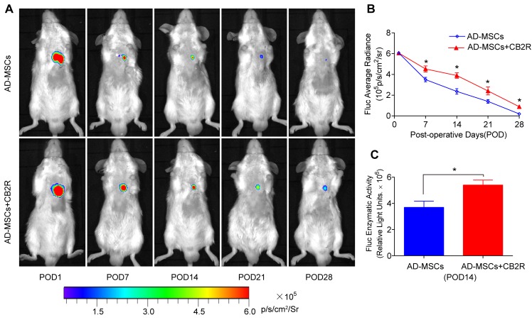
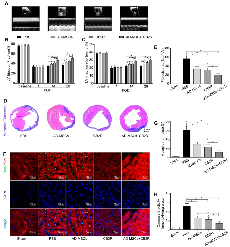
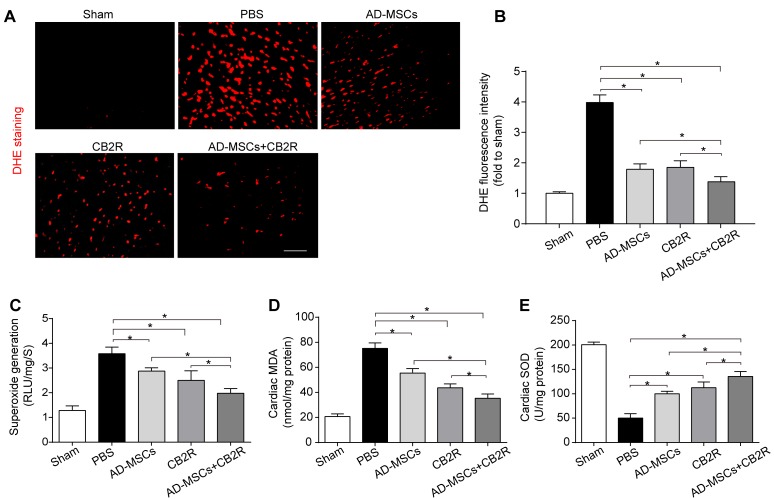
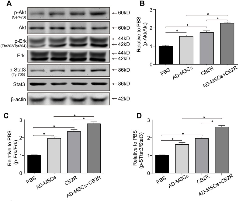
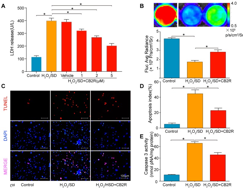
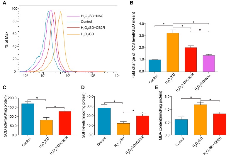
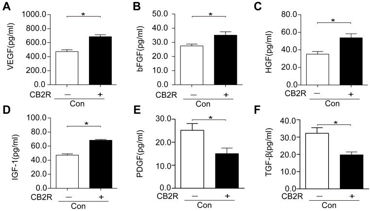
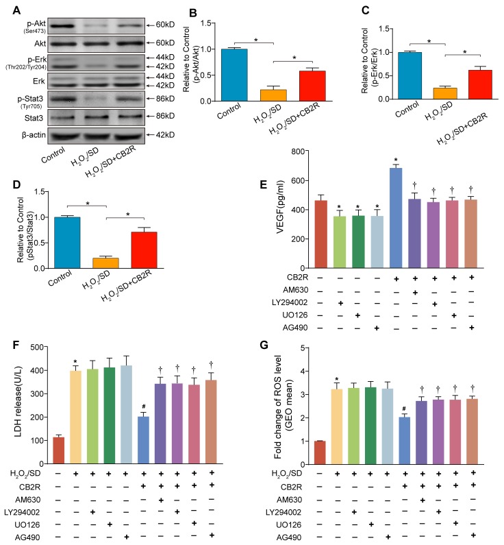
The poor survival of cells in ischemic sites diminishes the therapeutic efficacy of stem cell therapy. Previously we and others have reported that Cannabinoid receptor type II (CB2) is protective during heart ischemic injury for its anti-oxidative activity. However, whether CB2 activation could improve the survival and therapeutic efficacy of stem cells in ischemic myocardium and the underlying mechanisms remain elusive. Here, we showed evidence that CB2 agonist AM1241 treatment could improve the functional survival of adipose-derived mesenchymal stem cells (AD-MSCs) in vitro as well as in vivo. Moreover, AD-MSCs adjuvant with AM1241 improved cardiac function, and inhibited cardiac oxidative stress, apoptosis and fibrosis. To unveil possible mechanisms, AD-MSCs were exposed to hydrogen peroxide/serum deprivation to simulate the ischemic environment in myocardium. Results delineated that AM1241 blocked the apoptosis, oxidative damage and promoted the paracrine effects of AD-MSCs. Mechanistically, AM1241 activated signal transducers and activators of transcription 3 (Stat3) through the phosphorylation of Akt and ERK1/2. Moreover, the administration of AM630, LY294002, U0126 and AG490 (inhibitors for CB2, Akt, ERK1/2 and Stat3, respectively) could abolish the beneficial actions of AM1241. Our result support the promise of CB2 activation as an effective strategy to optimize stem cell-based therapy possibly through Stat3 activation.

Keywords: Pathology Section; Stat3; cannabinoid receptor type II; myocardial infarction; oxidative stress; stem cells.


Conflict of interest statement

CONFLICTS OF INTERESTS None.

Figures

Figure 1
Figure 1. CB2 agonist promoted survival of engrafted AD-MSCs in ischemic myocardium
A. Longitudinal BLI tracked the survival and retention of AD-MSCs survival in vivo (n = 8 for each group). Colored scale bar represents BLI radiance intensity in p/s/cm 2/sr. B. Quantitative analysis of A. C. Ex vivo Fluc enzymatic activity of infarcted myocardium on POD 14 (n = 5). . *P < 0.05 versus AD-MSCs.
Figure 2
Figure 2. Effects of AD-MSCs and CB2 agonist treatment on post-MI cardiac function, cardiac fibrosis and apoptosis
A. Representative M-mode images by echocardiography on POD28. Left ventricle ejection fraction B. and fractional shortening C. were calculated by M-mode echocardiography on baseline, POD 1, 14, and 28(n = 6). *P < 0.05 between indicated groups. D., E. Myocardial fibrosis was determined by Masson’s trichrome staining (n = 6-8, bar = 1 mm). F. Representative TUNEL graphs depicting myocardial apoptosis on POD3. TUNEL-positive cells (green fluorescence), cTnI (red fluorescence), and DAPI-positive nuclei (blue fluorescence), bar = 50μm; G. Graphs summarize apoptosis index calculated by the number of TUNEL-positive nuclei per 100 nuclei from five randomly selected fields and H. Myocardial caspase-3 activity (n = 6-8) was measured. *P < 0.05 between indicated groups.
Figure 3
Figure 3. CB2 agonist adjuvant with AD-MSCs leads to attenuation of myocardial oxidative stress
A., B. Representative images of dihydroethidium fluorescence staining that evaluated ROS generation in myocardium and bar graph summarizing fluorescence intensity on POD3(n = 5). bar = 100μm; C. Myocardial levels of O2- by lucigenin chemiluminescence method on POD3 (n = 5). D. Cardiac levels of MDA by enzyme-linked immunosorbent assay (ELISA) on POD3 (n = 5). E. Myocardial levels of SOD by testing kits. *P < 0.05 between indicated groups.
Figure 4
Figure 4. CB2 agonist adjuvant with AD-MSCs leads to myocardial activation of Stat3
A. Representative photographs of immune blot of Akt, Erk, Stat3 and their phosphorylation. (n = 3), B.-D. Quantitative analysis of A.. *P < 0.05 between indicated groups.
Figure 5
Figure 5. CB2 agonist attenuates H2O2/SD-induced AD-MSCs injury and apoptosis
A. LDH release in AD-MSCs after respective treatment. B. in vitro BLI for AD-MSCs viability. C. Quantitative analysis of B., n = 3. C. Representative photographs of TUNEL-stained AD-MSCs indicating cell apoptosis. D. Quantitative analysis of C., n = 6. E. Casepase3 activity in AD-MSCs, n = 6. *P < 0.05 between indicated groups.
Figure 6
Figure 6. CB2 agonist decreased oxidative stress levels in AD-MSCs subjected to H2O2/SD
A. Flow cytometry analysis of ROS production in AD-MSCs after indicated treatment, n = 3. B. Quantitative analysis of A., C.-E. The SOD, GSH, and MDA level determined by respective commercially available assay kit. n = 3-6. *P < 0.05 between indicated groups.
Figure 7
Figure 7. CB2 agonist enhanced the production of paracrine growth factors and inhibited profibrotic cytokines in AD-MSCs
(A-F) Paracrine growth factors including VEGF, bFGF, HGF, and IGF-1 as well as profibrotic cytokines including PDGF andTGFβ were examined by respective ELISA kits. *P<0.05 between indicated groups..
Figure 8
Figure 8. Activation of Stat3 through the phosphorylation of Akt and Erk1/2 was required for CB2 agonist-elicited beneficial actions in AD-MSCs
A. Representative photographs of immune blot of Akt, Erk, Stat3 and their phosphorylation. (n = 3), B.-D. Quantitative analysis of A.. *P < 0.05 between indicated groups. AD-MSCs were pre-treated with CB2 agonist (5 μM, 12 h) in the presence or absence of AM630 (4μM, a CB2 inhibitor), U0126 (5μM, an Erk1/2 inhibitor), LY294002 (5μM, a PI3K/Akt inhibitor) or AG490 (a Stat3 inhibitor, 40μM) and subsequently underwent H2O2/SD insult. E. Paracrine growth factor VEGF secreted by AD-MSCs following indicated treatment, F. LDH release by AD-MSCs after indicated treatment, n = 3. G. Flow cytometry analysis of ROS production in AD-MSCs after indicated treatment, n = 3. n = 3. *P < 0.05 versus blank control, #P < 0.05 versus H2O2/SD, P < 0.05 versus H2O2/SD+CB2R or CB2R.

References

    1. Garbern JC, Lee RT. Cardiac stem cell therapy and the promise of heart regeneration. Cell Stem Cell. 2013;12:689–698. - PMC - PubMed
    1. Abdelwahid E, Kalvelyte A, Stulpinas A, de Carvalho KA, Guarita-Souza LC, Foldes G. Stem cell death and survival in heart regeneration and repair. Apoptosis. 2016;21:252–268. - PMC - PubMed
    1. Golpanian S, Wolf A, Hatzistergos KE, Hare JM. Rebuilding the Damaged Heart: Mesenchymal Stem Cells, Cell-Based Therapy, and Engineered Heart Tissue. Physiol Rev. 2016;96:1127–1168. - PMC - PubMed
    1. Turcotte C, Blanchet MR, Laviolette M, Flamand N. The CB2 receptor and its role as a regulator of inflammation. Cell Mol Life Sci. 2016;73:4449–4470. - PMC - PubMed
    1. Scholl A, Ivanov I, Hinz B. Inhibition of interleukin-1β-induced endothelial tissue factor expression by the synthetic cannabinoid WIN 55,212-2. Oncotarget. 2016;7:61438–61457. doi: 10.18632/oncotarget.11367. - DOI - PMC - PubMed

LinkOut - more resources