. 2017 May 22;8(39):65186-65198.
doi: 10.18632/oncotarget.18053. eCollection 2017 Sep 12.

Serine 207 phosphorylated lysyl-tRNA synthetase predicts disease-free survival of non-small-cell lung carcinoma

Affiliations

Serine 207 phosphorylated lysyl-tRNA synthetase predicts disease-free survival of non-small-cell lung carcinoma

Suliman Boulos et al. Oncotarget. .

Erratum in

Abstract

It has been shown that various tRNA synthetases exhibit non-canonical activities unrelated to their original role in translation. We have previously described a signal transduction pathway in which serine 207 phosphorylated lysyl-tRNA synthetase (P-s207 LysRS) is released from the cytoplasmic multi-tRNA synthetase complex (MSC) into the nucleus, where it activates the transcription factor MITF in stimulated cultured mast cells and cardiomyocytes. Here we describe a similar transformation of LysRS due to EGFR signaling activation in human lung cancer. Our data shows that activation of the EGFR results in phosphorylation of LysRS at position serine 207, its release from the MSC and translocation to the nucleus. We then generated a P-s207 LysRS rabbit polyclonalantibody and tested 242 tissue micro-array samples derived from non-small-cell lung cancer patients. Highly positive nuclear staining for P-s207 LysRS was noted in patients with EGFR mutations as compared to WT EGFR patients and was associated with improved mean disease-free survival (DFS). In addition, patients with mutated EGFR and negative lymph node metastases had better DFS when P-s207 LysRS was present in the nucleus. The data presented strongly suggests functional and prognostic significance of P-s207 LysRS in non-small-cell lung cancer.

Keywords: EGFR; LysRS; P-s207 LysRS; multi-synthetase complex; non-small-cell lung cancer.


Conflict of interest statement

CONFLICTS OF INTEREST The authors have declared that no conflicts of interest exist.

Figures

Figure 1
Figure 1. EGFR-dependent LysRS release from the multi-synthetase complex (MSC) and translocation to the nucleus in NSCLC
(A) A549 cells were treated with 100 ng/ml of EGF for the indicated times. Phosphorylation of P-s207 LysRS was determined by immunoblotting using the rabbit polyclonal anti 207pLysRS antibody (B) Competitive ELISA with P-s207 LysRS antibody. P-s207 and wild type LysRS antigens (the peptides EITLLS (p) PCLHM and EITLLSPCLHM, respectively) were used at various concentrations as soluble competitors for P-s207 LysRS (C) Nuclear fractionation assay to determine the nuclear levels of P-s207 LysRS in A549 cells. To determine the localization of P-s207 LysRS, A549 cells were starved for 2 hr and incubated with EGF before nuclear and cytoplasmic fractionation. P-s207 LysRS levels were determined by immunoblotting. (D) Inhibition of ERK Phosphorylation by U0126 treatment in A549 and HCC827 cell lines. A549 cells were starved for 2hr and treated with EFG (100 ng/ml) for 15 min before exposure to U0126 (10 μM) for 10 min. HCC827 cells were treated with U0126 only. Cells were lysed and levels of P-s207 LysRS were determined by western blotting.
Figure 2
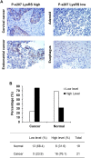
Figure 2. P-s207 LysRS levels in different tumor and normal tissue samples
(A) Examples taken from the original immunohistochemistry (IHC) slides showing high and low staining levels of P-s207 LysRS. (B) Summary of the IHC study showing in percentage the staining of P-s207 LysRS in tumors and normal tissue.
Figure 3
Figure 3. P-s207 LysRS levels in NSCLC tissues
(A) Scoring system for cytoplasmic staining of P-s207 LysRS in NSCLC tissues. The levels of cytoplasmic P-s207 LysRS staining were classified into 4 groups (0; none or positive in less than 10% of tumor cells, 1; weak positive in more than 10% of tumor cells 2; moderate positive in more than 10% of tumor cells 3; strong positive in more than 10% of tumor cells). (B) Scoring for nuclear P-s207 LysRS staining in NSCLC tissues, showing no staining (left figure) vs. high staining (right figure). Cases were deemed positive for nuclear P-s207 LysRS if more than 5% of tumor cells show nuclear staining of any intensity. (C) Validation of P-s207 LysRS antibody in IHC staining. To validate P-s207 LysRS-specific signal in IHC, P-s207 LysRS and LysRS antigens (that were used to produce the antibodies) were pre-incubated with the antibodies. (i) Control staining with the P-s207 LysRS antibody, (ii) P-s207 LysRS staining after pre-incubated with P-s207 LysRS antigen, (iii) P-s207 LysRS staining after pre-incubation with the LysRS antigen.
Figure 4
Figure 4. Nuclear P-s207 LysRS level is significantly associated with longer disease free survival (DFS) in NSCLC patients with mutated EGFR
Disease free survival of wild-type (A) and mutated EGFR patients (B) with (positive) or without (negative) nuclear P-s207 LysRS. Disease free survival of wild-type (C) and mutated EGFR patients (D) with (positive) or without (negative) nuclear LysRS.
Figure 5
Figure 5. Cytosolic and nuclear LysRS levels are not associated with longer disease free survival (DFS) in NSCLC patients with mutated EGFR
Disease free survival of wild-type EGFR patients with (positive) or without (negative) cytosolic (A) and nuclear (B) LysRS. Disease free survival of mutated EGFR patients with (positive) or without (negative) cytosolic (C) and nuclear (D) LysRS.
Figure 6
Figure 6. P-s207 LysRS level induces tumorigenicity
(A) Cells were transfected with WT LysRS, s207A and s207D LysRS mutant before colony formation assay. Cells were seeded on soft agar for 8 days then fluorescent absorption was detected at 485/520 nm wavelength. Results were analyzed by T-test. Table represents 6 independent experiments. (B) Transfectant expression level was determined by western blotting. Error bars represent the mean ± SEM. *P < 0.05; **P < 0.01.
Figure 7
Figure 7. Proposed model for the EGFR-LysRS signaling pathway in lung cancer
Following EGFR activation upon EGF binding, ERK is phosphorylated and activated. This results in the specific phosphorylation of LysRS at position Serine 207, its release from the MSC and translocation to the nucleus.

References

    1. Bandyopadhyay AK, Deutscher MP. Complex of aminoacyl-transfer RNA synthetases. J Mol Biol. 1971;60:113–122. - PubMed
    1. Guo M, Schimmel P. Essential nontranslational functions of tRNA synthetases. Nat Chem Biol. 2013;9:145–153. - PMC - PubMed
    1. Guo M, Yang XL, Schimmel P. New functions of aminoacyl-tRNA synthetases beyond translation. Nat Rev Mol Cell Biol. 2010;11:668–674. - PMC - PubMed
    1. Yannay-Cohen N, Carmi-Levy I, Kay G, Yang CM, Han JM, Kemeny DM, Kim S, Nechushtan H, Razin E. LysRS serves as a key signaling molecule in the immune response by regulating gene expression. Mol Cell. 2009;34:603–611. - PubMed
    1. Lee YN, Nechushtan H, Figov N, Razin E. The function of lysyl-tRNA synthetase and Ap4A as signaling regulators of MITF activity in FcepsilonRI-activated mast cells. Immunity. 2004;20:145–151. - PubMed