Review
. 2019 Jan 1:1702:74-84.
doi: 10.1016/j.brainres.2017.09.033. Epub 2017 Oct 10.

The heart of PD: Lewy body diseases as neurocardiologic disorders

Affiliations
Review

The heart of PD: Lewy body diseases as neurocardiologic disorders

David S Goldstein et al. Brain Res. .

Abstract

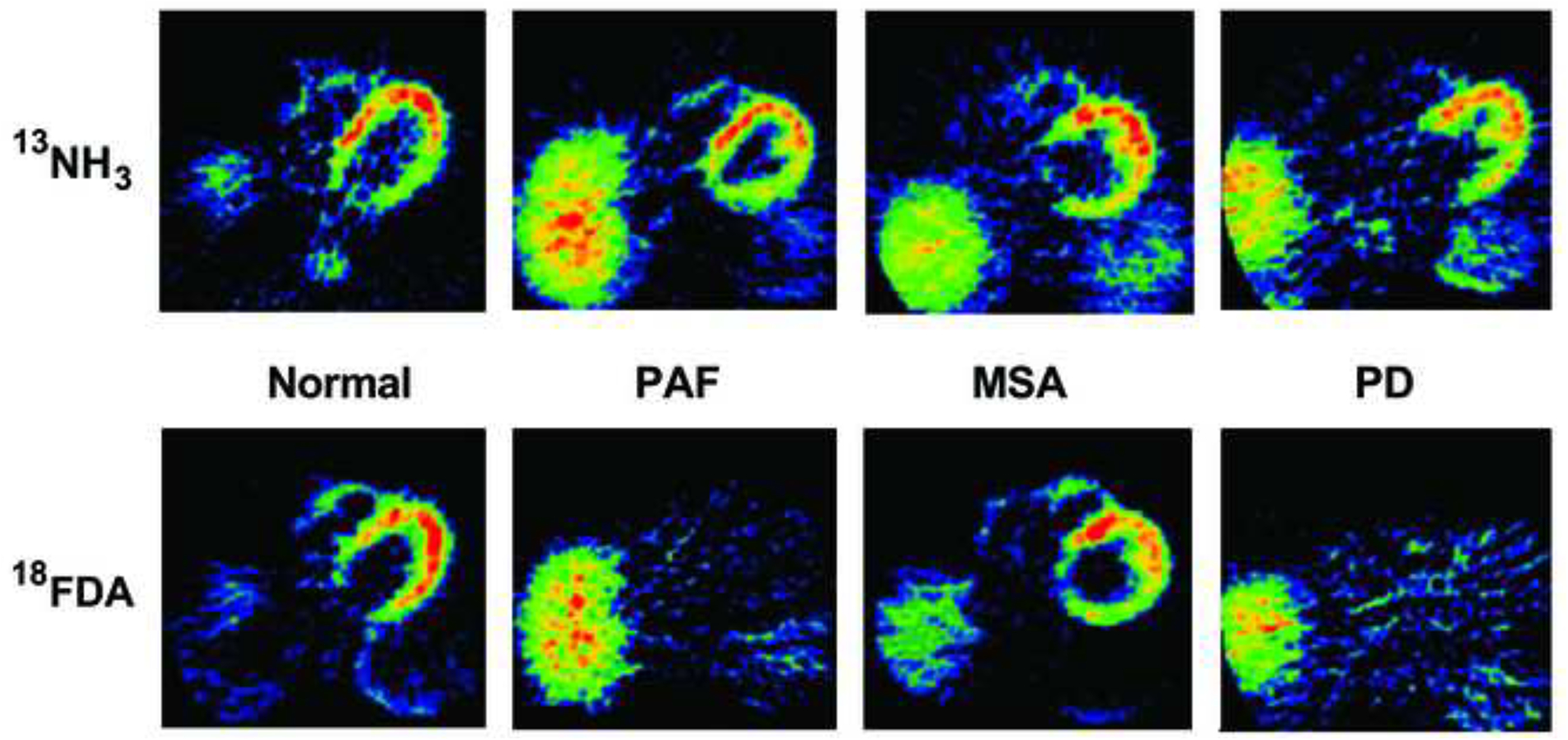
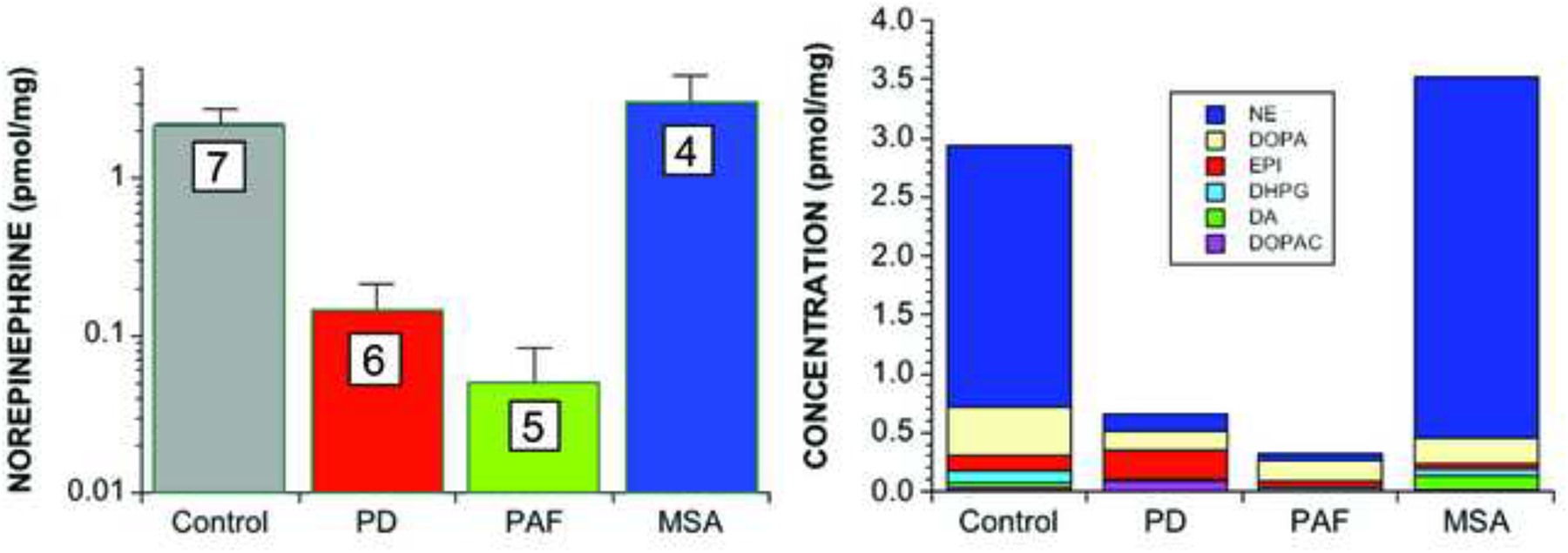
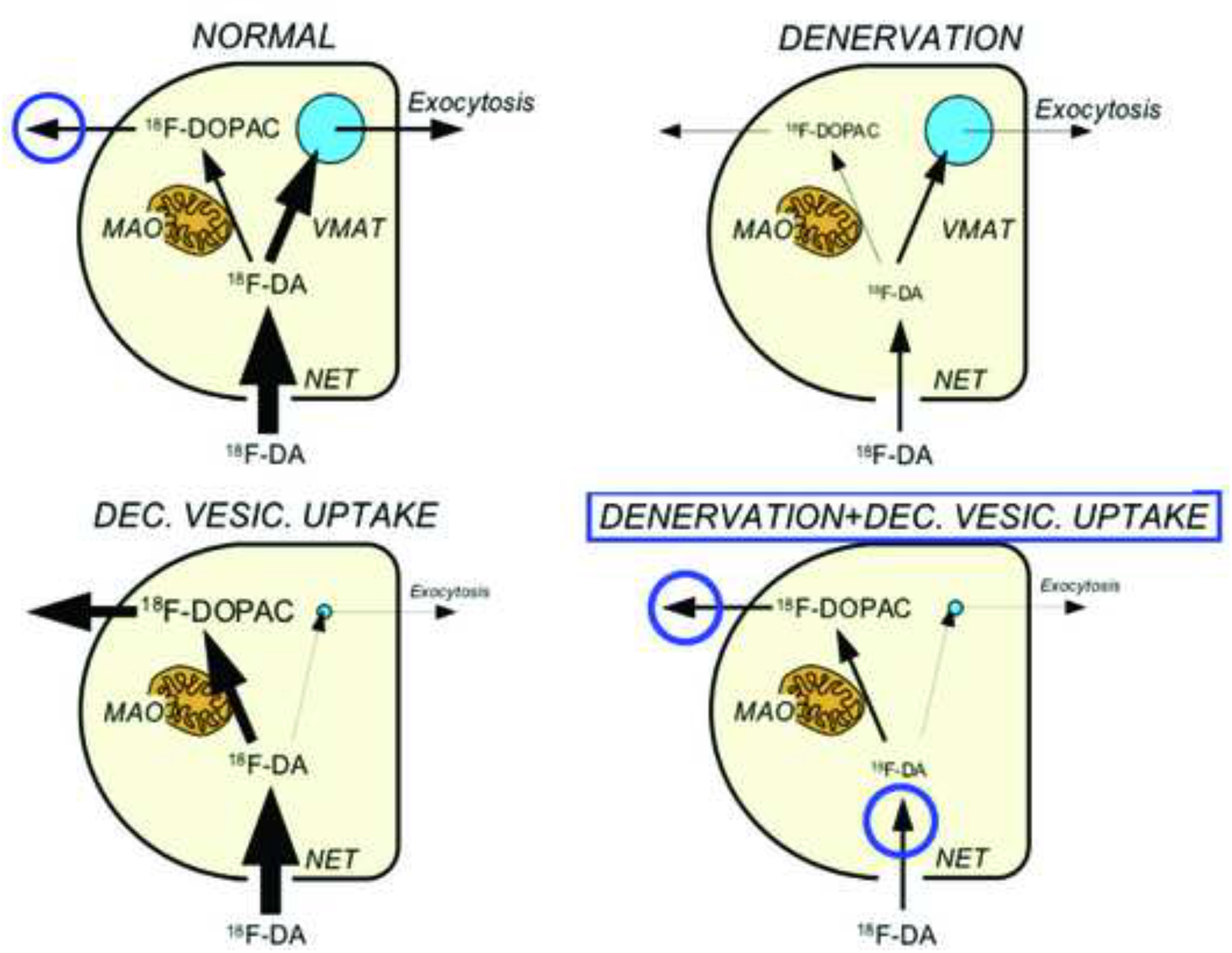
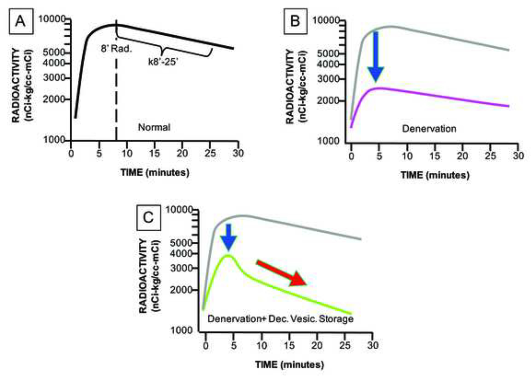
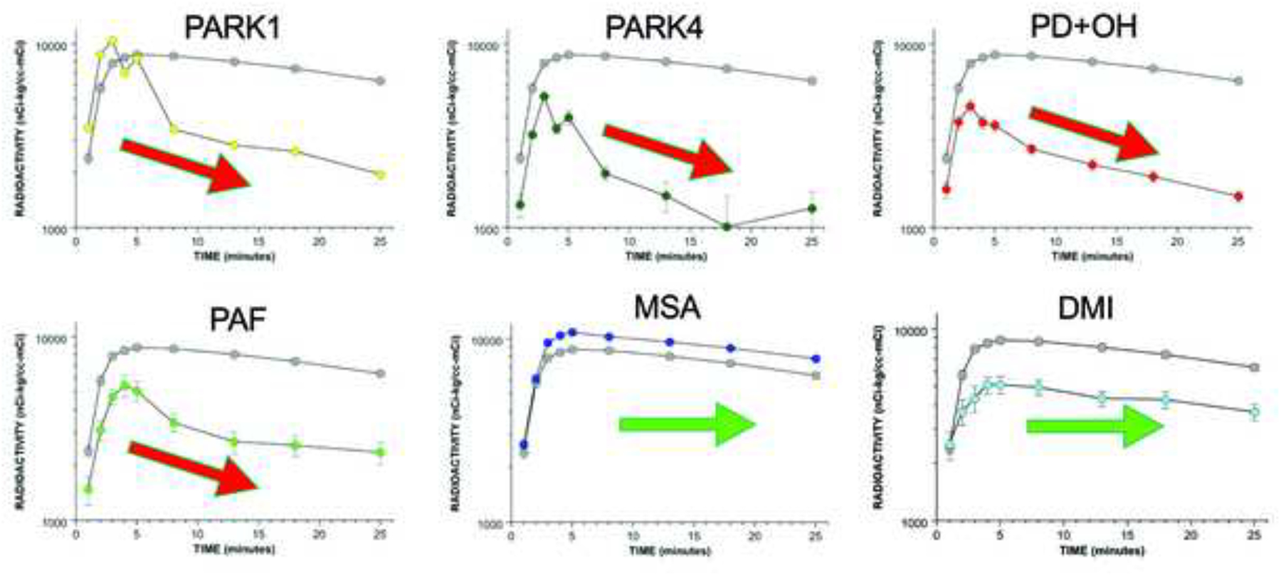
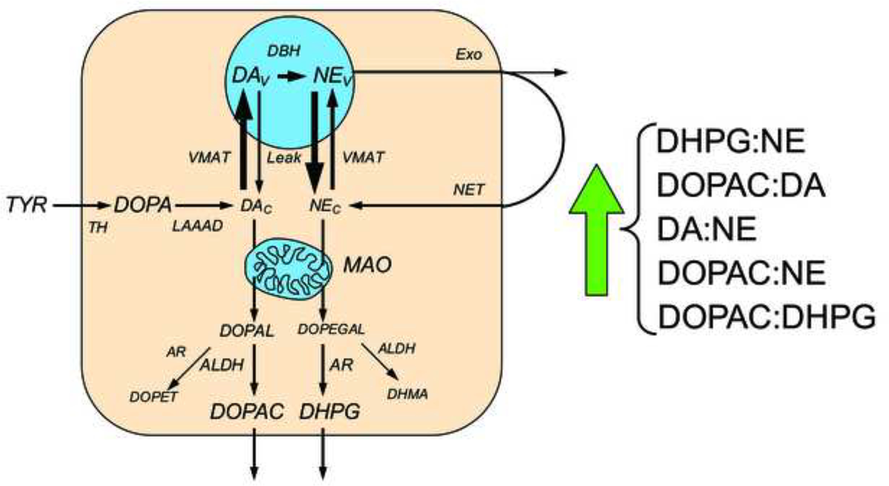
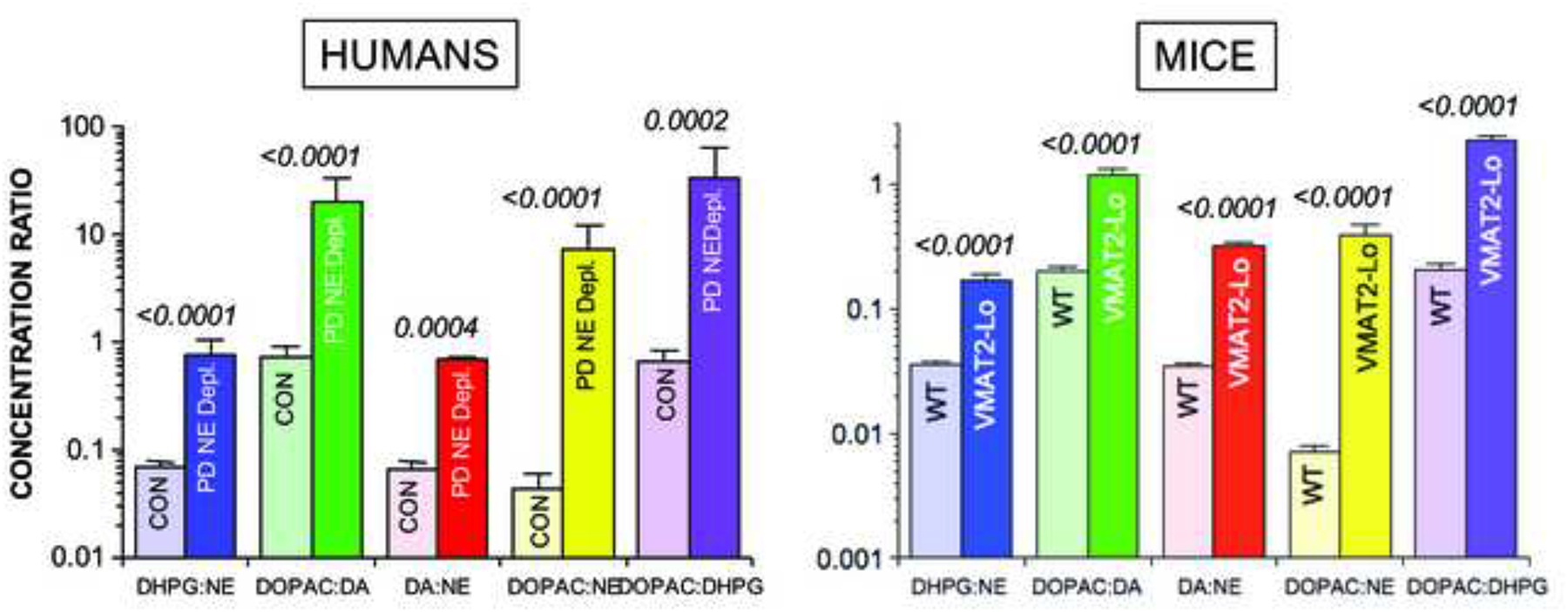
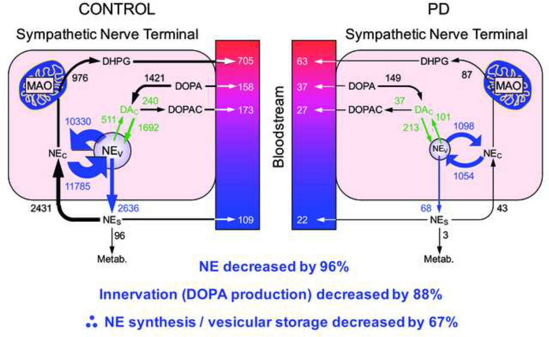
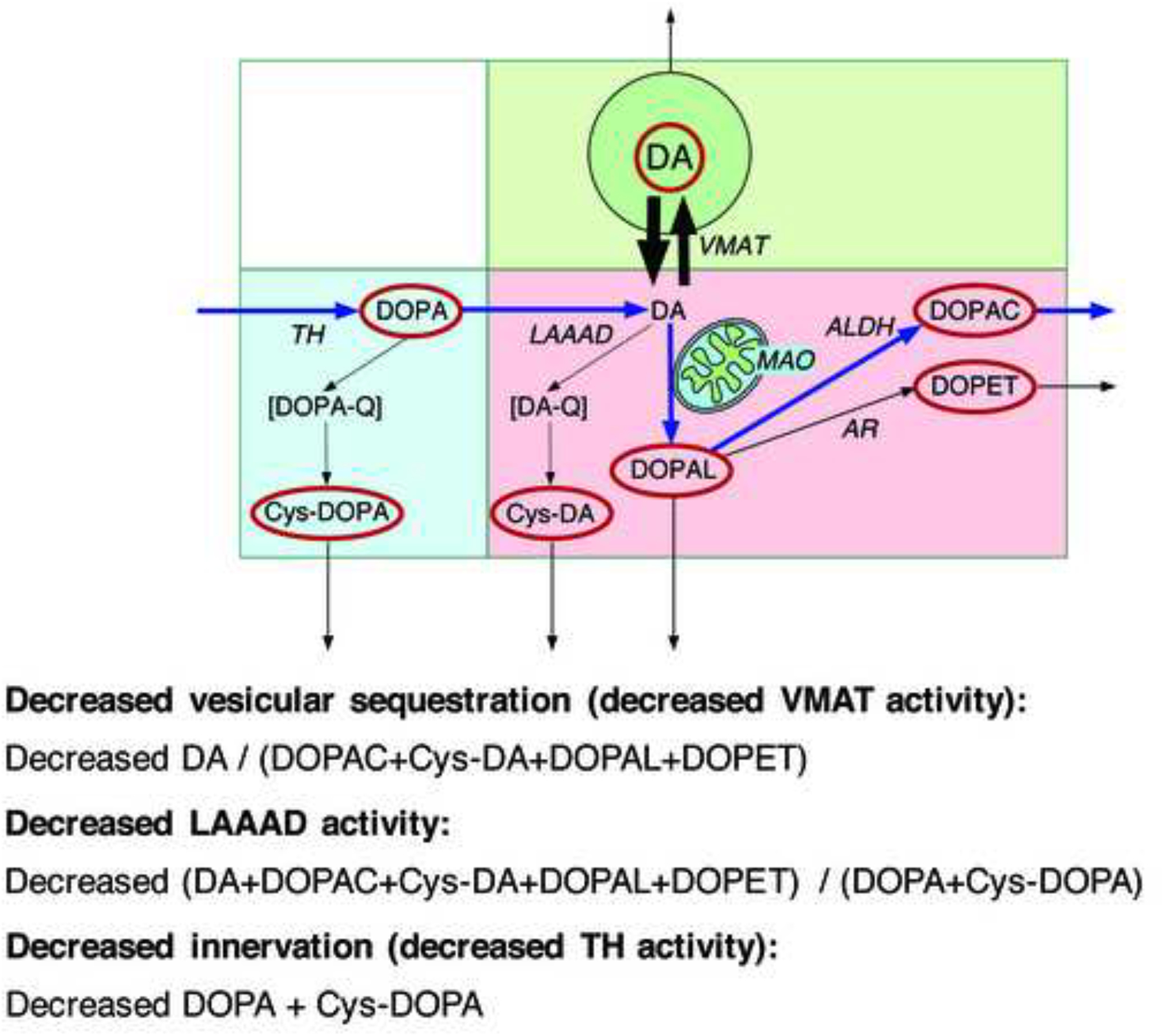
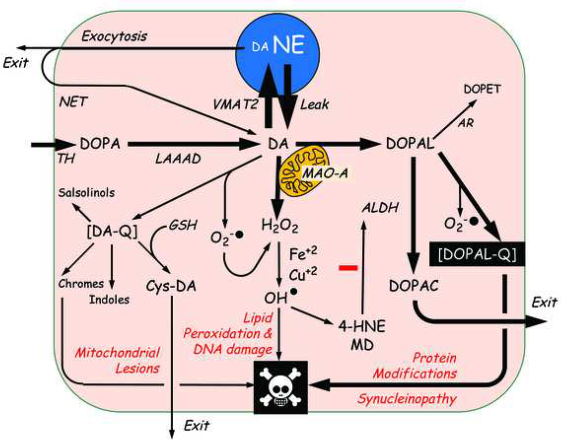
This review provides an update about cardiac sympathetic denervation in Lewy body diseases. The family of Lewy body diseases includes Parkinson's disease (PD), pure autonomic failure (PAF), and dementia with Lewy bodies (DLB). All three feature intra-neuronal cytoplasmic deposits of the protein, alpha-synuclein. Multiple system atrophy (MSA), the parkinsonian form of which can be difficult to distinguish from PD with orthostatic hypotension, involves glial cytoplasmic inclusions that contain alpha-synuclein. By now there is compelling neuroimaging, neuropathologic, and neurochemical evidence for cardiac sympathetic denervation in Lewy body diseases. In addition to denervation, there is decreased storage of catecholamines in the residual terminals. The degeneration develops in a centripetal, retrograde, "dying back" sequence. Across synucleinopathies the putamen and cardiac catecholaminergic lesions seem to occur independently of each other, whereas non-motor aspects of PD (e.g., anosmia, dementia, REM behavior disorder, OH) are associated with each other and with cardiac sympathetic denervation. Cardiac sympathetic denervation can be caused by synucleinopathy in inherited PD. According to the catecholaldehyde hypothesis, 3,4-dihydroxyphenylacetaldehyde (DOPAL), an intermediary metabolite of dopamine, causes or contributes to the death of catecholamine neurons, especially by interacting with proteins such as alpha-synuclein. DOPAL oxidizes spontaneously to DOPAL-quinone, which probably converts alpha-synuclein to its toxic oligomeric form. Decreasing DOPAL production and oxidation might slow the neurodegenerative process. Tracking cardiac sympathetic innervation over time could be the basis for a proof of principle experimental therapeutics trial targeting DOPAL.

Keywords: DOPAL; Dopamine; Norepinephrine; Parkinson’s disease; Sympathetic nervous system.


Conflict of interest statement

CONFLICT OF INTEREST

The authors have no conflicts of interest to disclose.

Figures

Figure 1:
Figure 1:. 13N-Ammonia and 18F-dopamine thoracic positron emission tomographic images from a normal control subject, a patient with pure autonomic failure (PAF), a patient with multiple system atrophy (MSA), and a patient with Parkinson’s disease (PD).
Note markedly decreased myocardial 18F-dopamine-derived radioactivity in PAF and PD.
Figure 2:
Figure 2:. Myocardial tissue contents of (left) norepinephrine and (right) 6 endogenous catechols in PD, PAF, and MSA.
Numbers in white boxes show the numbers of patients. Note large decreases in norepinephrine and the sum of endogenous catechols in PD and PAF.
Figure 3:
Figure 3:. Concept diagram showing effects of denervation, decreased vesicular uptake, and combined denervation and decreased vesicular uptake on myocardial 18F-dopamine-derived radioactivity and arterial plasma 18F-dihydroxyphenylacetic acid (18F-DOPAC).
Circles placed to indicate increased arterial 18F-DOPAC/18F-dopamine-derived radioactivity as an inverse index of vesicular uptake.
Figure 4:
Figure 4:. Concept diagram showing effects of denervation, and combined denervation and decreased vesicular uptake on myocardial 18F-dopamine-derived radioactivity.
Denervation should decrease peak initial radioactivity (8’ Rad.), and decreased vesicular uptake should increase the slope of decline of radioactivity (k8’-25’).
Figure 5:
Figure 5:. Mean (± SEM) interventricular septal myocardial concentrations of 18F-dopamine-derived radioactivity in groups with synucleinopathies or desipramine (DMI)-treated control subjects.
PARK1, PARK4, PD with orthostatic hypotension (PD+OH), and PAF are associated with accelerated declines in radioactivity, whereas MSA and DMI treatment are not.
Figure 6:
Figure 6:. Concept diagram about sources and metabolic fates of catecholamines in myocardial sympathetic nerves.
Under resting conditions, loss of norepinephrine (NE) from the neurons is due mainly to passive leakage from the vesicles (NEv) into the cytosol (NEc), followed by enzymatic deamination catalyzed by monoamine oxidase (MAO). Cytosolic NE is taken up into the vesicles via the type 2 vesicular monoamine transporter (VMAT). Release by exocytosis from the vesicles, with escape of reuptake via the cell membrane NE transporter (NET), is a minor determinant of NE turnover. NE loss is balanced by catecholamine biosynthesis from the action of cytosolic L-aromatic-amino-acid decarboxylase (LAAAD) on 3,4-dihydroxyphenylalanine (DOPA) produced from tyrosine (TYR) by tyrosine hydroxylase (TH) and of dopamine-beta-hydroxylase (DBH), which is localized in the vesicles. The action of MAO on cytosolic DA produces 3,4-dihydroxyphenylacetaldehyde (DOPAL) and on NE produces 3,4-dihydroxyphenylglycolaldehyde (DOPEGAL). DOPEGAL is mainly reduced by aldehyde/aldose reductase (AR), to form 3,4-dihydroxyphenylglycol (DHPG), and DOPAL is mainly oxidized by aldehyde dehydrogenase (ALDH) to form 3,4-dihydroxyphenylacetic acid (DOPAC). When vesicular uptake is attenuated, as in VMAT2-Lo mice, myocardial NE depletion reflects decreased NE synthesis, because less DA is taken up into the vesicles and more is deaminated to form DOPAC. Furthermore, decreased reuptake of NE that leaks from vesicles into the cytoplasm, where the NE is deaminated and is converted to DHPG, accelerates the turnover of NE. Decreased vesicular sequestration of cytoplasmic catecholamines would be expected to produce 5 alterations in ratios of catechols in myocardial tissue.
Figure 7:
Figure 7:. Mean (± SEM) ratios of catechols in myocardial tissue from (left) PD patients with norepinephrine depletion (NE Depl.) and control subjects (CON) and (right) mice with very low VMAT2 activity (VMAT2-Lo) and wild type (WT) mice.
Note similar abnormalities of indices of vesicular storage in PD patients with norepinephrine depletion and in VMAT-2 Lo mice.
Figure 8:
Figure 8:. Estimated rates of processes in myocardial sympathetic nerves in control subjects and in PD patients.
Norepinephrine (NE) depletion reflects both denervation (decreased DOPA production) and decreased vesicular storage in the residual sympathetic nerves. The model cannot distinguish decreased vesicular uptake from increased vesicular leakage and decreased intra-vesicular NE synthesis as determinants of tissue NE depletion.
Figure 9:
Figure 9:. Concept diagram about sources and metabolic fate of dopamine in putamen tissue.
Tyrosine hydroxylase (TH) catalyzes the conversion of tyrosine to DOPA, and L-aromatic-amino-acid decarboxylase (LAAAD) converts DOPA to dopamine (DA). Most of the DA in putamen tissue is in vesicles, due to uptake mediated by the vesicular monoamine transporter (VMAT). Cytoplasmic DA can be metabolized by monoamine oxidase (MAO) in the outer mitochondrial membrane to form 3,4-dihydroxyphenylacetaldehyde (DOPAL), which is metabolize by aldehyde dehydrogenase (ALDH) to form 3,4-dihydroxyphenylacetic acid (DOPAC) or by aldehyde/aldose reductase (AR) to form 3,4-dihydroxyphenylethanol (DOPET). Cytoplasmic DA can oxidize spontaneously to form DA-quinone (DA-Q) and then 5-S-cysteinyl-DA (Cys-DA), and cytoplasmic DOPA can oxidize spontaneously to form DOPA-quinone (DOPA-Q) and then 5-S-cysteinyl-DOPA (Cys-DOPA). The rectangle in aqua corresponds to products of TH proximal to DA; in pink to cytoplasmic DA metabolites; and in green to vesicular DA.
Figure 10:
Figure 10:. Myocardial 18F-dopamine-derived radioactivity as a function of time in (top) a PD patient with initially normal radioactivity and (bottom) a PD patient with decreased radioactivity in the apex and free wall.
In the PD patient at the top, radioactivity was normal for about 8 years, then free wall radioactivity declined, and then septal radioactivity followed after about 2 years. In the PD patient at the bottom, there was rapid loss of radioactivity over a few years. Red circles placed to indicate hypothesized slowing of cardiac sympathetic denervation in an experimental therapeutics trial.
Figure 11:
Figure 11:. Overview of the catecholamine autotoxicity theory as applied to cardiac sympathetic denervation in Lewy body diseases.
The theory explains PD in terms of toxic effects of products of enzymatic and spontaneous oxidation of cytoplasmic dopamine (DA), via mitochondrial lesions; lipid peroxidation and DNA damage, and protein modifications including alpha-synucleinopathy. The complex inter-relationships set the stage for allostatic load and multiple positive feedback loops that threaten neuronal integrity. Not shown are cytoplasmic norepinephrine (NE), its deamination via monoamine oxidase A (MAO-A) to form the catecholaldehyde 3,4-dihydroxyphenylglycolaldehyde (DOPEGAL), and the reduction of DOPEGAL via aldehyde reductase (AR) to form 3,4-dihydroxyphenylglycol (DHPG). Other abbreviations: ALDH=aldehyde dehydrogenase; Cys-DA=5-S-cysteinyldopamine; DA-Q=dopamine quinone; DOPAC=3,4-dihydroxyphenylacetic acid; DOPAL=3,4-dihydroxyphenylacetaldehyde; DOPAL-Q=DOPAL quinone; GSH=reduced glutathione; 4-HNE=4-hydroxynonenal; LAAAD=L-aromatic-amino-acid decarboxylase; MD=malondialdehyde; NET=cell membrane norepinephrine transporter; TH=tyrosine hydroxylase; VMAT2=type 2 vesicular monoamine transporter.

References

    1. Anderson DG, Florang VR, Schamp JH, Buettner GR, Doorn JA, 2016. Antioxidant-mediated modulation of protein reactivity for 3,4-dihydroxyphenylacetaldehyde, a toxic dopamine metabolite. Chem. Res. Toxicol 29, 1098–107. - PMC - PubMed
    1. Braak H, Ghebremedhin E, Rub U, Bratzke H, Del Tredici K, 2004. Stages in the development of Parkinson’s disease-related pathology. Cell Tissue Res. 318, 121–134. - PubMed
    1. Burke WJ, Li SW, Williams EA, Nonneman R, Zahm DS, 2003. 3,4-Dihydroxyphenylacetaldehyde is the toxic dopamine metabolite in vivo: implications for Parkinson’s disease pathogenesis. Brain Res. 989, 205–13. - PubMed
    1. Burke WJ, Kumar VB, Pandey N, Panneton WM, Gan Q, Franko MW, O’Dell M, Li SW, Pan Y, Chung HD, Galvin JE, 2008. Aggregation of alpha-synuclein by DOPAL, the monoamine oxidase metabolite of dopamine. Acta Neuropathol. 115, 193–203. - PubMed
    1. Chu Y, Morfini GA, Langhamer LB, He Y, Brady ST, Kordower JH, 2012. Alterations in axonal transport motor proteins in sporadic and experimental Parkinson’s disease. Brain. 135, 2058–73. - PMC - PubMed

Publication types

MeSH terms