

Impact of peripheral immune status on central molecular responses to facial nerve axotomy
- PMID: 29030217
- PMCID: PMC5767532
- DOI: 10.1016/j.bbi.2017.10.005
Impact of peripheral immune status on central molecular responses to facial nerve axotomy
Abstract
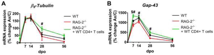
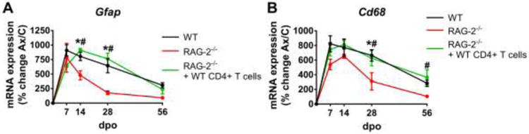
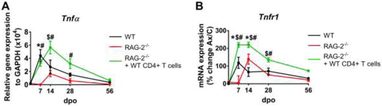
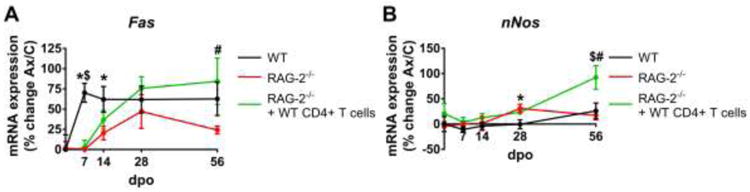
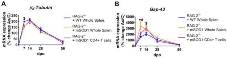
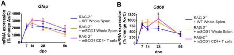
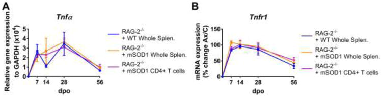
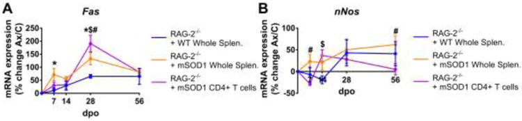
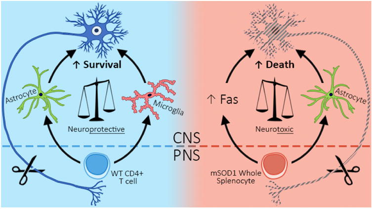
When facial nerve axotomy (FNA) is performed on immunodeficient recombinase activating gene-2 knockout (RAG-2-/-) mice, there is greater facial motoneuron (FMN) death relative to wild type (WT) mice. Reconstituting RAG-2-/- mice with whole splenocytes rescues FMN survival after FNA, and CD4+ T cells specifically drive immune-mediated neuroprotection. Evidence suggests that immunodysregulation may contribute to motoneuron death in amyotrophic lateral sclerosis (ALS). Immunoreconstitution of RAG-2-/- mice with lymphocytes from the mutant superoxide dismutase (mSOD1) mouse model of ALS revealed that the mSOD1 whole splenocyte environment suppresses mSOD1 CD4+ T cell-mediated neuroprotection after FNA. The objective of the current study was to characterize the effect of CD4+ T cells on the central molecular response to FNA and then identify if mSOD1 whole splenocytes blocked these regulatory pathways. Gene expression profiles of the axotomized facial motor nucleus were assessed from RAG-2-/- mice immunoreconstituted with either CD4+ T cells or whole splenocytes from WT or mSOD1 donors. The findings indicate that immunodeficient mice have suppressed glial activation after axotomy, and cell transfer of WT CD4+ T cells rescues microenvironment responses. Additionally, mSOD1 whole splenocyte recipients exhibit an increased astrocyte activation response to FNA. In RAG-2-/- + mSOD1 whole splenocyte mice, an elevation of motoneuron-specific Fas cell death pathways is also observed. Altogether, these findings suggest that mSOD1 whole splenocytes do not suppress mSOD1 CD4+ T cell regulation of the microenvironment, and instead, mSOD1 whole splenocytes may promote motoneuron death by either promoting a neurotoxic astrocyte phenotype or inducing Fas-mediated cell death pathways. This study demonstrates that peripheral immune status significantly affects central responses to nerve injury. Future studies will elucidate the mechanisms by which mSOD1 whole splenocytes promote cell death and if inhibiting this mechanism can preserve motoneuron survival in injury and disease.
Keywords: Amyotrophic lateral sclerosis; Axotomy; Motoneuron; Nerve injury; T cells.
Copyright © 2017 Elsevier Inc. All rights reserved.
Figures
Similar articles
-
SOD1(G93A) transgenic mouse CD4(+) T cells mediate neuroprotection after facial nerve axotomy when removed from a suppressive peripheral microenvironment.Brain Behav Immun. 2014 Aug;40:55-60. doi: 10.1016/j.bbi.2014.05.019. Epub 2014 Jun 6. Brain Behav Immun. 2014. PMID: 24911596 Free PMC article.
-
Axotomy-induced target disconnection promotes an additional death mechanism involved in motoneuron degeneration in amyotrophic lateral sclerosis transgenic mice.J Comp Neurol. 2014 Jul 1;522(10):2349-76. doi: 10.1002/cne.23538. J Comp Neurol. 2014. PMID: 24424947 Free PMC article.
-
CD4 + T Cells and Neuroprotection: Relevance to Motoneuron Injury and Disease.J Neuroimmune Pharmacol. 2015 Dec;10(4):587-94. doi: 10.1007/s11481-015-9625-x. Epub 2015 Jul 7. J Neuroimmune Pharmacol. 2015. PMID: 26148561 Free PMC article. Review.
-
CD4(+) T cell-mediated facial motoneuron survival after injury: Distribution pattern of cell death and rescue throughout the extent of the facial motor nucleus.J Neuroimmunol. 2006 Dec;181(1-2):93-9. doi: 10.1016/j.jneuroim.2006.08.006. Epub 2006 Oct 11. J Neuroimmunol. 2006. PMID: 17045343
-
Role of the immune system in the maintenance of mouse facial motoneuron viability after nerve injury.Brain Behav Immun. 2005 Jan;19(1):12-9. doi: 10.1016/j.bbi.2004.05.004. Brain Behav Immun. 2005. PMID: 15581733 Review.
Cited by
-
Temporospatial Analysis and New Players in the Immunology of Amyotrophic Lateral Sclerosis.Int J Mol Sci. 2018 Feb 23;19(2):631. doi: 10.3390/ijms19020631. Int J Mol Sci. 2018. PMID: 29473876 Free PMC article. Review.
-
CD4+ T cell expression of the IL-10 receptor is necessary for facial motoneuron survival after axotomy.J Neuroinflammation. 2020 Apr 17;17(1):121. doi: 10.1186/s12974-020-01772-x. J Neuroinflammation. 2020. PMID: 32303238 Free PMC article.
-
Neurodegenerative diseases and neuroinflammation-induced apoptosis.Open Life Sci. 2025 Feb 25;20(1):20221051. doi: 10.1515/biol-2022-1051. eCollection 2025. Open Life Sci. 2025. PMID: 40026360 Free PMC article. Review.
References
-
- Alexianu ME, Kozovska M, Appel SH. Immune reactivity in a mouse model of familial ALS correlates with disease progression. Neurology. 2001;57(7):1282–1289. - PubMed
-
- Aloisi F, Ria F, Columba-Cabezas S, Hess H, Penna G, Adorini L. Relative efficiency of microglia, astrocytes, dendritic cells and B cells in naive CD4+ T cell priming and Th1/Th2 cell restimulation. Eur J Immunol. 1999;29(9):2705–2714. doi: 10.1002/(SICI)1521-4141(199909)29:09<2705∷AID-IMMU2705>3.0.CO;2-1. - DOI - PubMed
Publication types
MeSH terms
Substances
Grants and funding
LinkOut - more resources
Full Text Sources
Other Literature Sources
Molecular Biology Databases
Research Materials
Miscellaneous
