. 2017 Oct;10(5):e001770.
doi: 10.1161/CIRCGENETICS.117.001770.

Transcriptome and Functional Profile of Cardiac Myocytes Is Influenced by Biological Sex

Affiliations

Transcriptome and Functional Profile of Cardiac Myocytes Is Influenced by Biological Sex

Christa L Trexler et al. Circ Cardiovasc Genet. 2017 Oct.

Abstract

Background: Although cardiovascular disease is the primary killer of women in the United States, women and female animals have traditionally been omitted from research studies. In reports that do include both sexes, significant sexual dimorphisms have been demonstrated in development, presentation, and outcome of cardiovascular disease. However, there is little understanding of the mechanisms underlying these observations. A more thorough understanding of sex-specific cardiovascular differences both at baseline and in disease is required to effectively consider and treat all patients with cardiovascular disease.

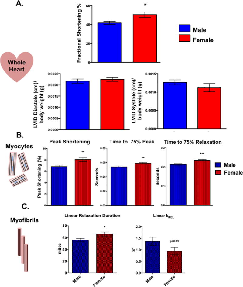
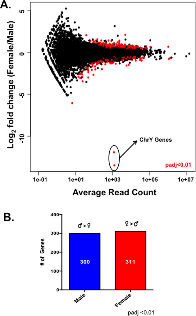
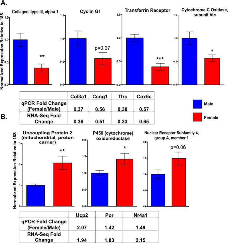
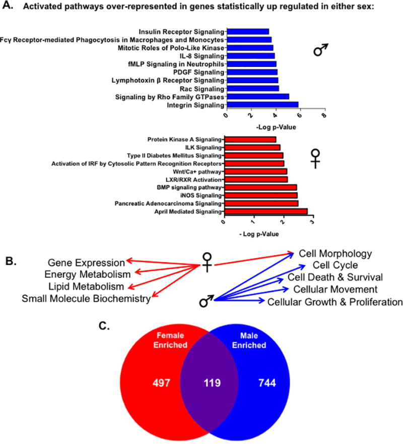
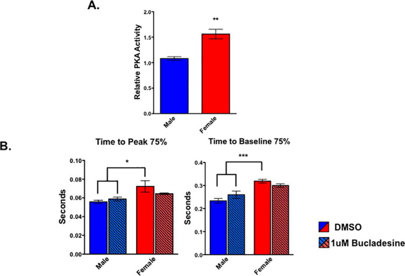
Methods and results: We analyzed contractility in the whole rat heart, adult rat ventricular myocytes (ARVMs), and myofibrils from both sexes of rats and observed functional sex differences at all levels. Hearts and ARVMs from female rats displayed greater fractional shortening than males, and female ARVMs and myofibrils took longer to relax. To define factors underlying these functional differences, we performed an RNA sequencing experiment on ARVMs from male and female rats and identified ≈600 genes were expressed in a sexually dimorphic manner. Further analysis revealed sex-specific enrichment of signaling pathways and key regulators. At the protein level, female ARVMs exhibited higher protein kinase A activity, consistent with pathway enrichment identified through RNA sequencing. In addition, activating the protein kinase A pathway diminished the contractile sexual dimorphisms previously observed.

Conclusions: These data support the notion that sex-specific gene expression differences at baseline influence cardiac function, particularly through the protein kinase A pathway, and could potentially be responsible for differences in cardiovascular disease presentation and outcomes.

Keywords: cardiovascular diseases; gene expression; myocytes, cardiac; rats; sequence analysis, RNA; sex differences.


Figures

Figure 1
Figure 1. Cardiac function is sexually dimorphic from the level of the whole heart down to the myofibril
(A) In vivo whole heart function as measured by echocardiography in male and female rats. *p<0.05 relative to male, N=5 female and 6 males. (B) Contractile function of electrically paced ventricular myocytes from rats of both sex. **p<0.01, ***p<0.001 relative to male, N=97 female cells and 110 male cells pooled from 6 animals of each sex. (C) Kinetic properties of left ventricular myofibrils isolated from male and female rats. Linear relaxation duration: *p<0.05 relative to female, N=21 female and 25 male myofibrils from 3 separate animals. Linear KREL: N= 15 female and 14 myofibrils from 3 separate animals. All data reported as mean ± SEM.
Figure 2
Figure 2. Male and female cardiac myocytes exhibit different transcriptome profiles
(A) Differential gene expression analysis between male and female cardiac myocytes as measured with DESeq. Differentially expressed genes between the sexes are indicated by the red circles; padj <0.01 female relative to male, N=5 female and 6 male isolated cardiac myocyte preparations. (B) The number of differentially expressed genes (padj<0.01) genes at least 1.5× higher in one sex relative to the other as measured by DESeq; this list of genes was used for downstream analysis with IPA.
Figure 3
Figure 3. Validation of differentially expressed genes with qPCR also demonstrates that cardiac myocyte gene expression is sexually dimorphic
Expression of Col3a1, Ccng1, Tfrc, Cox6c (A), Ucp2, Por, and Nr4a1 (B) as measured by qPCR in male and female cardiac myocytes. The mean fold changes (female relative to male) for each gene as found by qPCR and the RNA-sequencing experiment are indicated in the supplied tables. All genes were normalized to levels of 18S; *p<0.05, **p<0.01, ***p<0.001 female relative to male; N=5 female and 6 male isolated cardiac myocyte preparations. All data reported as mean ± SEM.
Figure 4
Figure 4. Specific pathways and functions are enriched in cardiac myocytes from either sex
IPA analysis of significantly expressed genes by at least 1.5 fold in either sex. (A) The top ten statistically significant canonical pathways predicted to be activated by IPA in genes unregulated in either male or female cardac myocytes. Pathways were deemed statistically significant by IPA if p<0.05 using the Fischer exact test. Activated pathways were indicated by positive z-scores calculated by IPA; for pathways that contained the same genes, the pathway with the lower p-value was listed. (B) The top five statistically significant (lower end of p-value overlap at least 0.05) molecular and cellular functions enriched in genes found to be upregulated in either male or female myocytes by IPA. (C) The number of statistically significant (p<0.05) upstream regulators enriched in up-regulated genes from either sex as measured by IPA analysis.
Figure 5
Figure 5. PKA activity is sexually dimorphic in cardiac myocytes
(A) Levels of PKA activity in male and female cardiac myocytes as measured by PKA activity assay. **p<0.01 female relative to male; n=4 animals of each sex. All data reported as mean ± SEM. (B) Contractile function of electrically paced ventricular myocytes treated with bucladesine or DMSO for 30 minutes from rats of both sex. *p<0.05 relative to female DMSO; n= at least 19 cells per group pooled from 4 animals of each sex.

Comment in

  • Sex Determines Cardiac Myocyte Stretch and Relaxation.
    Coronado MJ, Fairweather D, Bruno KA. Coronado MJ, et al. Circ Cardiovasc Genet. 2017 Oct;10(5):e001950. doi: 10.1161/CIRCGENETICS.117.001950. Circ Cardiovasc Genet. 2017. PMID: 29030406 Free PMC article. No abstract available.

References

    1. Mozaffarian D, Benjamin EJ, Go AS, Arnett DK, Blaha MJ, Cushman M, et al. Heart disease and stroke statistics-2015 update: A report from the american heart association. Circulation. 2015;131:e29–322. - PubMed
    1. Miller VM, Best PJ. Implications for reproductive medicine: Sex differences in cardiovascular disease. Sex Reprod Menopause. 2011;9:21–28. - PMC - PubMed
    1. Consideration of sex as a biological variable in nih-funded research. 2015
    1. Clayton JA, Collins FS. Policy: Nih to balance sex in cell and animal studies. Nature. 2014;509:282–283. - PMC - PubMed
    1. Regitz-Zagrosek V. Therapeutic implications of the gender-specific aspects of cardiovascular disease. Nat Rev Drug Discov. 2006;5:425–438. - PubMed

MeSH terms