

Infliximab for the treatment of CNS sarcoidosis: A multi-institutional series
- PMID: 29030454
- PMCID: PMC5711506
- DOI: 10.1212/WNL.0000000000004644
Infliximab for the treatment of CNS sarcoidosis: A multi-institutional series
Abstract
Objective: To describe clinical and imaging responses in neurosarcoidosis to infliximab, a monoclonal antibody against tumor necrosis factor-α.
Methods: Investigators at 6 US centers retrospectively identified patients with CNS sarcoidosis treated with infliximab, including only patients with definite or probable neurosarcoidosis following rigorous exclusion of other causes.
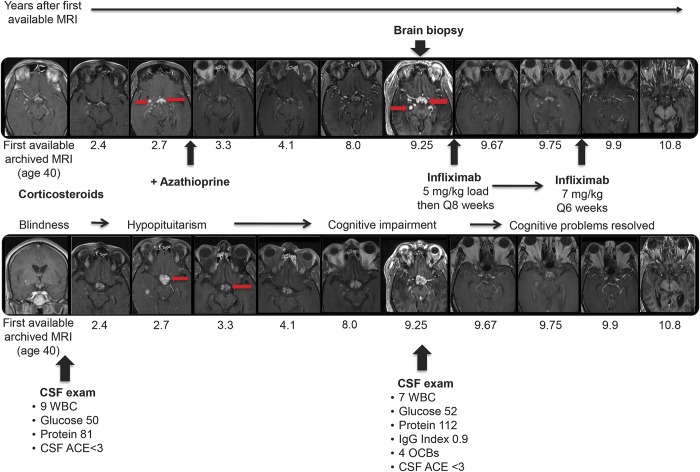
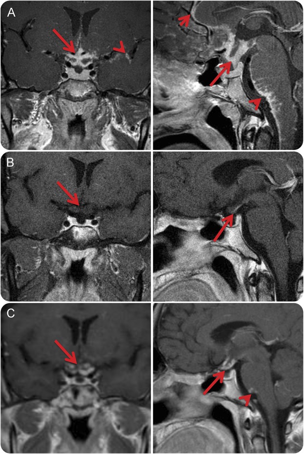
Results: Of 66 patients with CNS sarcoidosis (27 definite, 39 probable) treated with infliximab for a median of 1.5 years, the mean age was 47.5 years at infliximab initiation (SD 11.7, range 24-71 years); 56.1% were female; 62.1% were white, 37.0% African American, and 3% Hispanic. Sarcoidosis was isolated to the CNS in 19.7%. Using infliximab doses ranging from 3 to 7 mg/kg every 4-8 weeks, MRI evidence of a favorable treatment response was observed in 82.1% of patients with imaging follow-up (n = 56), with complete remission of active disease in 51.8% and partial MRI improvement in 30.1%; MRI worsened in 1 patient (1.8%). There was clinical improvement in 77.3% of patients, with complete neurologic recovery in 28.8%, partial improvement in 48.5%, clinical stability in 18.2%, worsening in 3%, and 1 lost to follow-up. In 16 patients in remission when infliximab was discontinued, the disease recurred in 9 (56%), typically in the same neuroanatomic location.
Conclusions: Most patients with CNS sarcoidosis treated with infliximab exhibit favorable imaging and clinical treatment responses, including some previously refractory to other immunosuppressive treatments.
Classification of evidence: This study provides Class IV evidence that for patients with CNS sarcoidosis infliximab is associated with favorable imaging and clinical responses.
© 2017 American Academy of Neurology.
Figures
Comment in
-
Reader response: Infliximab for the treatment of CNS sarcoidosis: A multi-institutional series.Neurology. 2018 Jul 3;91(1):51. doi: 10.1212/WNL.0000000000005727. Neurology. 2018. PMID: 29967207 No abstract available.
-
Author response: Infliximab for the treatment of CNS sarcoidosis: A multi-institutional series.Neurology. 2018 Jul 3;91(1):51-52. doi: 10.1212/WNL.0000000000005725. Neurology. 2018. PMID: 29967208 No abstract available.
References
-
- Iannuzzi MC, Rybicki BA, Teirstein AS. Sarcoidosis. N Engl J Med 2007;357:2153–2165. - PubMed
-
- Rossi G, Cavazza A, Colby TV. Pathology of sarcoidosis. Clin Rev Allergy Immunol 2015;49:36–44. - PubMed
-
- Fingerlin TE, Hamzeh N, Maier LA. Genetics of sarcoidosis. Clin Chest Med 2015;36:569–584. - PubMed
-
- Rybicki BA, Iannuzzi MC. Epidemiology of sarcoidosis: recent advances and future prospects. Semin Respir Crit Care Med 2007;28:22–35. - PubMed
-
- Zajicek JP, Scolding NJ, Foster O, et al. . Central nervous system sarcoidosis: diagnosis and management. QJM 1999;92:103–117. - PubMed
MeSH terms
Substances
Supplementary concepts
Grants and funding
LinkOut - more resources
Full Text Sources
Other Literature Sources
Medical
Research Materials