

Conjugation with L,L-diphenylalanine Self-Assemblies Enhances In Vitro Antitumor Activity of Phthalocyanine Photosensitizer
- PMID: 29030603
- PMCID: PMC5640658
- DOI: 10.1038/s41598-017-13729-x
Conjugation with L,L-diphenylalanine Self-Assemblies Enhances In Vitro Antitumor Activity of Phthalocyanine Photosensitizer
Abstract
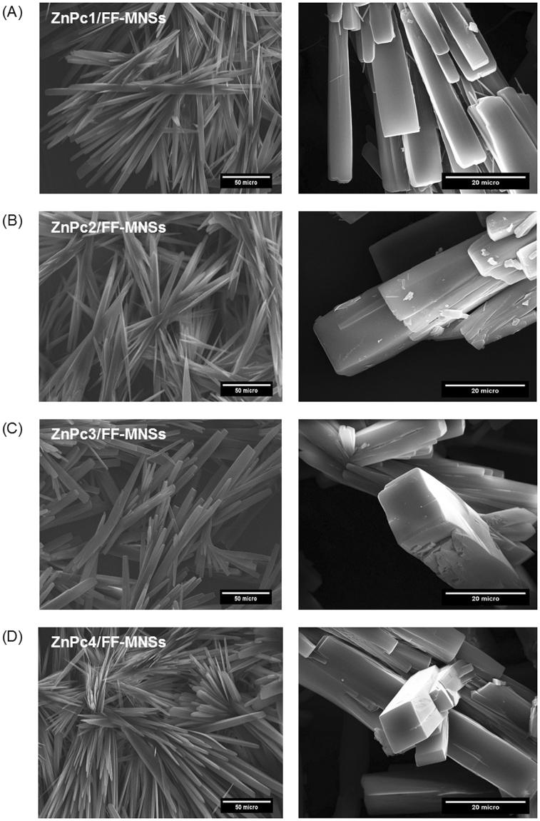
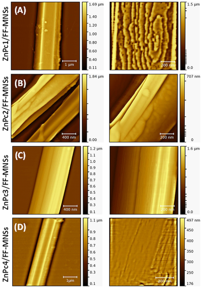
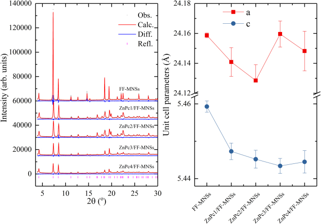
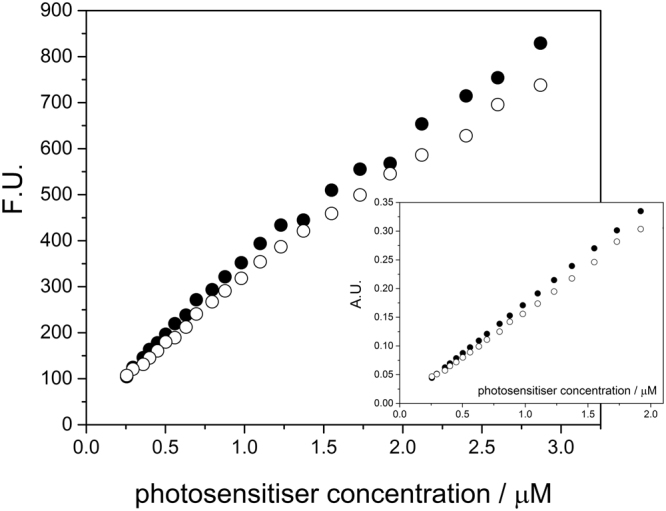
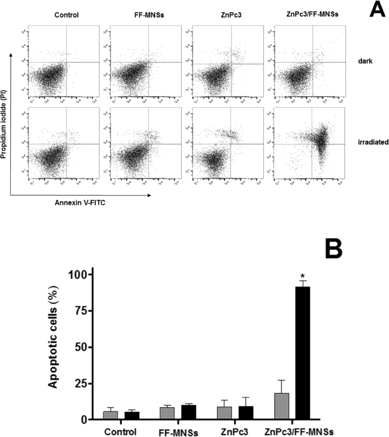
We present the synthesis and characterization of new peptide conjugates obtained by hierarchical co-assembly of L,L-diphenylalanine (FF) and zinc phthalocyanine complexes (ZnPc) in water. Self-assembly capabilities under defined conditions were investigated by scanning electron microscopy, and photophysical properties were evaluated using UV-Vis and fluorescence spectroscopy. AFM observations demonstrated that these ZnPcs form different highly ordered arrays on the crystalline faces of the FF microplates and that surface roughness significantly changes with the presence of differently substituted phthalocyanine units. XRD assays showed that the overall molecular packing of the conjugates is organized according to a hexagonal symmetry, with ZnPcs hosted in the interstices of the peptide phase. In vitro photodynamic studies were conducted on human breast cancer MCF-7 cells to investigate both cellular uptake and cytotoxicity. It was shown that FF self-assemblies are not toxicity and enhance accumulation of ZnPc in MCF-7 cells, improving apoptotic cell death upon irradiation. Our findings demonstrate enhancement of ZnPc antitumor efficiency by FF conjugates and a proof-of-concept for new photosensitizer carriers based on peptide conjugates.
Conflict of interest statement
The authors declare that they have no competing interests.
Figures


Similar articles
-
Novel theranostic zinc phthalocyanine-phospholipid complex self-assembled nanoparticles for imaging-guided targeted photodynamic treatment with controllable ROS production and shape-assisted enhanced cellular uptake.Colloids Surf B Biointerfaces. 2018 Feb 1;162:76-89. doi: 10.1016/j.colsurfb.2017.10.061. Epub 2017 Oct 24. Colloids Surf B Biointerfaces. 2018. PMID: 29154189
-
Lenvatinib-zinc phthalocyanine conjugates as potential agents for enhancing synergistic therapy of multidrug-resistant cancer by glutathione depletion.Eur J Med Chem. 2019 May 1;169:53-64. doi: 10.1016/j.ejmech.2019.02.065. Epub 2019 Mar 4. Eur J Med Chem. 2019. PMID: 30856406
-
Novel zinc phthalocyanine as a promising photosensitizer for photodynamic treatment of esophageal cancer.Int J Oncol. 2017 Mar;50(3):953-963. doi: 10.3892/ijo.2017.3854. Epub 2017 Jan 17. Int J Oncol. 2017. PMID: 28098886
-
Tetra-triethyleneoxysulfonyl substituted zinc phthalocyanine for photodynamic cancer therapy.Photodiagnosis Photodyn Ther. 2016 Mar;13:148-157. doi: 10.1016/j.pdpdt.2015.07.001. Epub 2015 Jul 7. Photodiagnosis Photodyn Ther. 2016. PMID: 26162500 Review.
-
Pharmaceutical development, composition and quantitative analysis of phthalocyanine as the photosensitizer for cancer photodynamic therapy.J Pharm Biomed Anal. 2014 Jan;87:98-104. doi: 10.1016/j.jpba.2013.05.014. Epub 2013 May 18. J Pharm Biomed Anal. 2014. PMID: 23746989 Review.
Cited by
-
Synthesis, Characterization and Evaluation of Peptide Nanostructures for Biomedical Applications.Molecules. 2021 Jul 29;26(15):4587. doi: 10.3390/molecules26154587. Molecules. 2021. PMID: 34361740 Free PMC article. Review.
-
Peptide-Tetrapyrrole Supramolecular Self-Assemblies: State of the Art.Molecules. 2021 Jan 28;26(3):693. doi: 10.3390/molecules26030693. Molecules. 2021. PMID: 33525730 Free PMC article. Review.
References
-
- Bank-Srour B, et al. Physical vapor deposition of peptide nanostructures. Polym. J. 2013;45:494–503. doi: 10.1038/pj.2013.19. - DOI
Publication types
MeSH terms
Substances
LinkOut - more resources
Full Text Sources
Other Literature Sources
Miscellaneous
