. 2017 Oct 13;7(1):13149.
doi: 10.1038/s41598-017-13558-y.

Lubricin binds cartilage proteins, cartilage oligomeric matrix protein, fibronectin and collagen II at the cartilage surface

Affiliations

Lubricin binds cartilage proteins, cartilage oligomeric matrix protein, fibronectin and collagen II at the cartilage surface

Sarah A Flowers et al. Sci Rep. .

Abstract

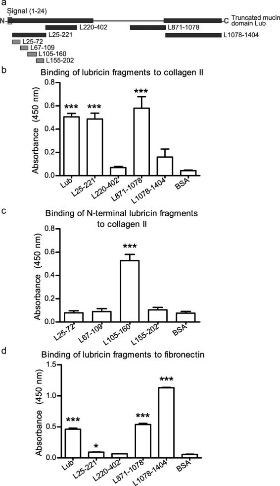
Lubricin, a heavily O-glycosylated protein, is essential for boundary lubrication of articular cartilage. Strong surface adherence of lubricin is required given the extreme force it must withstand. Disulfide bound complexes of lubricin and cartilage oligomeric matrix protein (COMP) have recently been identified in arthritic synovial fluid suggesting they may be lost from the cartilage surface in osteoarthritis and inflammatory arthritis. This investigation was undertaken to localise COMP-lubricin complexes within cartilage and investigate if other cartilage proteins are involved in anchoring lubricin to the joint. Immunohistochemical analysis of human cartilage biopsies showed lubricin and COMP co-localise to the cartilage surface. COMP knockout mice, however, presented with a lubricin layer on the articular cartilage leading to the further investigation of additional lubricin binding mechanisms. Proximity ligation assays (PLA) on human cartilage biopsies was used to localise additional lubricin binding partners and demonstrated that lubricin bound COMP, but also fibronectin and collagen II on the cartilage surface. Fibronectin and collagen II binding to lubricin was confirmed and characterised by solid phase binding assays with recombinant lubricin fragments. Overall, COMP, fibronectin and collagen II bind lubricin, exposed on the articular cartilage surface suggesting they may be involved in maintaining essential boundary lubrication.


Conflict of interest statement

The authors declare that they have no competing interests.

Figures

Figure 1
Figure 1
Immunohistochemical co-localisation of lubricin and COMP. Dual antibody immunofluorescence on OA cartilage biopsy cryosections with anti-lubricin (P3-118) and anti-COMP (mAb 16F12) antibodies. Section shows an area of undisrupted cartilage surface (left side) and an area more severely affected by OA including a tear (right side). (a) Haemotoxylin and esosin staining. (b) DAPI staining for nuclei of chondrocytes. (c) COMP (green) was constantly distributed over the section with greater intensity on the superficial zone. (d) Lubricin (red) was present on the superficial zone of the cartilage and into the superficial zone in the area of the tissue with OA degradation at the surface. (e) Cartilage surface alone including COMP antibody staining, lubricin antibody staining and merged image showing co-localisation of COMP and lubricin on cartilage surface. Negative controls of dual antibody immunofluorescence analysis. The specificity of the staining was verified using matched isotype negative controls or control serum at the same concentration as the primary antibodies. (f) Negative control showing DAPI staining. (g) Negative control for the anti-COMP antibody. (h) Negative control for the anti-lubricin antibody.
Figure 2
Figure 2
Immunohistochemical localisation of lubricin in WT and COMP KO mice paw joint cartilage tissue. (a) WT mouse tissue stained with rabbit anti-lubricin (P3-118) visualised with Rhodamine Red-X shows a diffuse lubricin layer at the cartilage surface. (b) COMP KO mouse tissue stained with rabbit anti-lubricin (P3-118) visualised with Rhodamine Red-X showing a discrete, distinct layer of lubricin at the cartilage surface. (c) WT mouse tissue stained with rabbit anti-lubricin (P3-118) visualised with Rhodamine Red-X also showing DAPI staining. (d) COMP KO mouse tissue stained with rabbit anti-lubricin (P3-118) visualised with Rhodamine Red-X also showing DAPI staining. (e) Negative control of WT mouse paw tissue performed by omitting primary antibody.
Figure 3
Figure 3
In situ PLA of lubricin with possible cartilage binding partners. In situ PLA was performed on OA cartilage biopsy cryosections with the following pairs. In situ PLA uses two primary antibodies targeting proteins of interest and secondary antibodies conjugated to DNA oligonucleotides (PLA probes). If the two proteins of interest are in a complex the two PLA probes will be in close enough proximity to bind and facilitate formation of a circular DNA molecule using two additional DNA oligonucleotides which then undergo rolling circle amplification to enhance the signal. Fluorescently (red) labelled complementary DNA is then added in order to visualise the DNA. (a) Lubricin (PA3-118) and COMP (16F12). Bound lubricin and COMP (red) were identified at the cartilage surface. A second layer was also apparent below the surface. (b) Lubricin (5C11) and fibronectin (ab32419). Bound lubricin and fibronectin (red) were predominately identified at the cartilage surface. Only when the PLA signal was thinner at the surface was signal observed just below the surface. (c) Lubricin (5C11) and collagen II (sc-7763). Bound lubricin and collagen II (red) were identified only at the surface with no PLA signal further into the tissue. PLA signal are red, DAPI staining in blue. For the 3 antibody pairs, 5 separate antibodies were used as anti-lubricin 5CII was used for 2 pairs. Negative controls were performed by omitting each primary antibody for all of the five antibodies used. (d) lubricin negative control (PA3-118), (e) COMP negative control (16F12), (f) lubricin negative control (5CII), (g) fibronectin negative control (ab32419) and (h) collagen II negative control (sc-7763).
Figure 4
Figure 4
Recombinant (RC) lubricin forms non-covalent complexes with collagen II and fibronectin. (a) Representation of RC lubricin constructs. Dark blue: FLAG-tagged, expressed in mammalian 293 F cells. Light blue: N-terminal fragments GST-tagged expressed in E. coli strain Rosetta 2. (b) Interaction between collagen II isolated from bovine cartilage and RC lubricin fragments, full length lubricin and BSA by solid phase binding assay. (c) Interaction between collagen II isolated from bovine cartilage and RC N-terminal lubricin fragments and BSA by solid phase binding assay. (d) Interaction between fibronectin isolated from human blood and RC lubricin fragments, full length lubricin and BSA by solid phase binding assay. For all assays n = 3 and error bars are standard deviation. N- designates the amino terminus and –C designates the carboxy terminus. The signal peptide (1–24) is shown in grey. All statistical analyses compare the binding of lubricin fragments to the binding of the BSA standard. * is defined as p ≤ 0.05, and *** is defined as p ≤ 0.001.
Figure 5
Figure 5
Western blots of lubricin released from the cartilage surface by MMP-9. (a) Cartilage biopsy tissue pieces were incubated with pro- and activated MMP-9 and released proteins were separated by SDS-PAGE and analysed by Western blot with lubricin antibody mAb13. (b) Purified lubricin was also incubated with activated MMP-9. Lubricin was released from the cartilage biopsy pieces when the surface of the cartilage was digested by MMP enzymes which digest the proteins of the cartilage ECM.
Figure 6
Figure 6
Illustration of the cartilage surface showing protein interactions. Lubricin is blue with glycosylation shown in light blue, COMP is red shown as the pentameric structure, fibronectin is green shown as the dimeric structure and collagen II is purple shown as collagen II fibrils. The SF is shown in light straw, the cartilage in yellow and the cartilage surface in pink. COMP, collagen II and fibronectin adhere lubricin, primarily by the termini of lubricin, to the cartilage surface creating an exposed lubricin layer. The ECM proteins also bind to each other and are shown in the cartilage. Lubricin is also found in the SF.

References

    1. Chen D, et al. Osteoarthritis: toward a comprehensive understanding of pathological mechanism. Bone research. 2017;5:16044. doi: 10.1038/boneres.2016.44. - DOI - PMC - PubMed
    1. Lee AS, et al. A current review of molecular mechanisms regarding osteoarthritis and pain. Gene. 2013;527:440–447. doi: 10.1016/j.gene.2013.05.069. - DOI - PMC - PubMed
    1. Smolen JS, Aletaha D, McInnes IB. Rheumatoid arthritis. Lancet. 2016;388:2023–2038. doi: 10.1016/S0140-6736(16)30173-8. - DOI - PubMed
    1. Rosman Z, Shoenfeld Y, Zandman-Goddard G. Biologic therapy for autoimmune diseases: an update. BMC medicine. 2013;11:88. doi: 10.1186/1741-7015-11-88. - DOI - PMC - PubMed
    1. Allen KD, et al. Osteoarthritis: Models for appropriate care across the disease continuum. Best practice & research. Clinical rheumatology. 2016;30:503–535. doi: 10.1016/j.berh.2016.09.003. - DOI - PubMed

Publication types