

Effects of regional perfusion block in healthy and injured lungs
- PMID: 29030751
- PMCID: PMC5640557
- DOI: 10.1186/s40635-017-0161-2
Effects of regional perfusion block in healthy and injured lungs
Abstract
Background: Severe hypoperfusion can cause lung damage. We studied the effects of regional perfusion block in normal lungs and in the lungs that had been conditioned by lavage with 500 ml saline and high V T (20 ml kg-1) ventilation.
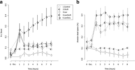
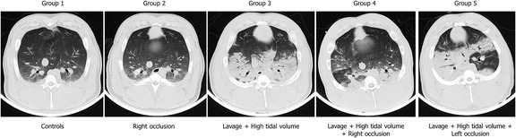
Methods: Nineteen pigs (61.2 ± 2.5 kg) were randomized to five groups: controls (n = 3), the right lower lobe block alone (n = 3), lavage and high V T (n = 4), lung lavage, and high V T plus perfusion block of the right (n = 5) or left (n = 4) lower lobe. Gas exchange, respiratory mechanics, and hemodynamics were measured hourly. After an 8-h observation period, CT scans were obtained at 0 and 15 cmH2O airway pressure.
Results: Perfusion block did not damage healthy lungs. In conditioned lungs, the left perfusion block caused more edema in the contralateral lung (777 ± 62 g right lung vs 484 ± 204 g left; p < 0.05) than the right perfusion block did (581 ± 103 g right lung vs 484 ± 204 g left; p n.s.). The gas/tissue ratio, however, was similar (0.5 ± 0.3 and 0.8 ± 0.5; p n.s.). The lobes with perfusion block were not affected (gas/tissue ratio right 1.6 ± 0.9; left 1.7 ± 0.5, respectively). Pulmonary artery pressure, PaO2/FiO2, dead space, and lung mechanics were more markedly affected in animals with left perfusion block, while the gas/tissue ratios were similar in the non-occluded lobes.
Conclusions: The right and left perfusion blocks caused the same "intensity" of edema in conditioned lungs. The total amount of edema in the two lungs differed because of differences in lung size. If capillary permeability is altered, increased blood flow may induce or increase edema.
Keywords: Computed tomography (CT); Experimental animal model; Lung injury; Pulmonary circulation; Pulmonary embolism; Ventilator-induced lung injury.
Conflict of interest statement
Ethics approval and consent to participate
The local authorities (Niedersächsisches Landesamt für Verbraucherschutz und Lebensmittelsicherheit LAVES; AZ 33.9–42,502–04-15/1757) approved the study.
Consent for publication
Not applicable.
Competing interests
The authors declare that they have no competing interests.
Publisher’s Note
Springer Nature remains neutral with regard to jurisdictional claims in published maps and institutional affiliations.
Figures

References
-
- Slutsky AS, Ranieri VM (2013) Ventilator-induced lung injury. N Engl J Med 369:2126–36 - PubMed
-
- Edmunds LH, Jr, Holm JC. Effect of atelectasis on lung changes after pulmonary arterial ligation. J Appl Physiol. 1968;25:115–123. - PubMed
-
- Edmunds LH, Jr, Holm JC. Effect of inhaled CO2 on hemorrhagic consolidation due to unilateral pulmonary arterial ligation. J Appl Physiol. 1969;26:710–715. - PubMed
-
- Kolobow T, Spragg RG, Pierce JE. Massive pulmonary infarction during total cardiopulmonary bypass in unanesthetized spontaneously breathing lambs. Int J Artif Organs. 1981;4:76–81. - PubMed
LinkOut - more resources
Full Text Sources
Other Literature Sources
Miscellaneous
